"Zoom" and modules
In the previous chapter I emphasized that the primary visual cortex appears morphologically uniform both to the naked eye and under the microscope using most conventional staining techniques. However, upon closer study, it turned out that the topography of the columns of ocular dominance is also homogeneous: the period of alternation of zones of dominance of the left and right eyes remains surprisingly constant from the projection of the central fovea (fixation point) to the far periphery of the binocular part of the visual field. Using the deoxyglucose injection method, we also revealed the uniformity of the topography of the orientation columns.
We initially perceived the fact of the anatomical homogeneity of the cortex as a surprise, since from a functional point of view this area is clearly heterogeneous in two important parameters. First, as discussed in Chapter 3, the receptive fields of retinal ganglion cells in and around the fovea are much smaller than those in the periphery. The dimensions of the receptive field of a typical complex cell in the upper layer of the cortex in the area of the projection of the fovea are approximately 0.25?0.5°. If we consider those parts of the cortex where the periphery of the retina is represented (80–90° from the fovea), then receptive fields measuring 2–4° are most often found here. If we compare the corresponding areas, the ratio between them can be estimated as 1:10 or even 1:30.
The second type of heterogeneity is associated with the so-called "increase". This concept, introduced in 1961 by P. Daniel and D. Whitteridge, refers to the distance between two points in the cortex to which two points of the visual field are projected, separated by a distance of 1°. If you go from the fovea to the periphery, the cortical projection of the same angular distance will become smaller and smaller, i.e. "increase" will decrease. If the stimulus is shifted by 1° near the fixation point, its projection in the cortex will shift by approximately 6 mm. If we place this stimulus in a zone 90° away from the fixation point, then a shift in the visual field of 1° will correspond to a shift along the surface of the cortex of only 0.15 mm. Thus, the "magnification" in the center of the retina is approximately 36 times greater than in the periphery.
Both considered inhomogeneities are associated with the same circumstance - with a decrease in visual acuity as one moves away from the center of the retina to its periphery. Try, for example, to fix your gaze on a letter at the very beginning of a line and try to determine which letter or word is at the very end. Or fix your gaze on the letter п at the beginning of a word progressive. It is unlikely that you will be able to identify the letter о at the end of a word. It will probably be difficult for you to identify в и н before the last letter. In order to provide high resolution in the fovea, the visual system requires many cortical cells per unit area in the projection of this region, and each such cell must have a very small receptive field.
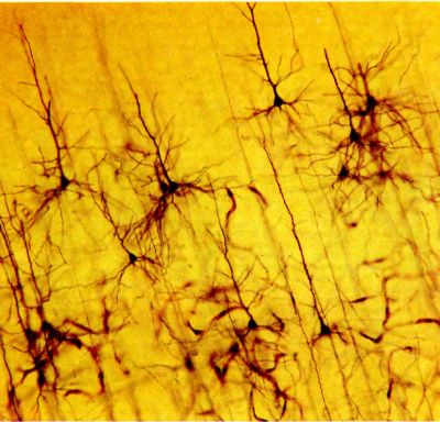
Rice. 90. This chapter will talk about “modules.” One module is roughly the size of the Golgi-stained section of visual cortex shown here. This staining method reveals only a very small proportion of all nerve cells, but if the cell is stained, then all or almost all of it is visible: in such a section you can see the cell body, its dendrites and axon.
Scatter and shift of receptive fields
Given the above, how can we explain the anatomical homogeneity of the cortex? To do this we will have to take a closer look at what? occurs with the position of the receptive fields of neurons encountered along the path of electrode advancement. If we insert an electrode into the striate cortex exactly at a right angle to its surface, it turns out that all these receptive fields are located almost in the same part of the visual field, but do not coincide completely - the position of the receptive field changes slightly from cell to cell. However, these changes appear to be random and not very large, so that the receptive fields of every two successive cells overlap significantly (this fact is reflected in Fig. 91). In any given cortical layer, the size of the receptive fields is relatively constant, but when moving to another layer they change noticeably - from very small in layer 4C to large in layers 5 and 6. Within one cortical layer, the area occupied by ten or twelve sequentially recorded fields, due to random scatter, will be approximately 2-4 times larger than the area of the receptive field of one cell. Let us name the area occupied by the entire set of receptive fields of individual cells in one layer and at one point of the cortex composite receptive field this point. Depending on the layer of the cortex, the magnitude of the composite field varies - for example, in layer 3 it is about 30 arc minutes for the central fovea and about 7–8° for the far periphery.
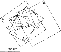
Rice. 91. Mapping of these nine receptive fields was carried out by passing a microelectrode once through the cat's striate cortex perpendicular to its surface. With such advancement of the electrode, random deviations in the position of the receptive fields are observed, as well as in their magnitude, but no tendency towards their systematic displacement is noted.
Now suppose that we insert and advance the electrode horizontally within one layer of the cortex, say layer 3. And here, when sequentially recording the responses of the cells, we will find that the position of the receptive fields varies randomly, but in these random shifts we will notice a tendency to shift in a certain direction. Of course, this direction can be predicted based on the topographic projection of the visual field into the cortex. Now we will be interested in the amount of displacement that is observed when the electrode is shifted by 1 millimeter horizontally. Considering what has been said above regarding differences in "magnification", it is clear that the distance measured in the field of view will depend on where exactly in the cortex we advance the electrode - in the area of the fovea, in the peripheral area, or somewhere in between. The rate of displacement of receptive fields in the visual field will be far from the same. It turns out, however, that this speed will be very close to constant if we correct for the size of the receptive fields themselves. A displacement anywhere in the cortex by 1 millimeter corresponds to a shift in the visual field that is equal to approximately half the diameter of the composite receptive field (i.e., the total field formed by the superposition of the receptive fields of all cells located under a given point on the surface of the cortex. Thus, in order to completely leave one part of the visual field and move to another, the next one, a displacement of approximately 2 millimeters will be required (this is shown in Fig. 92). The same turns out to be true for all points of the field 17, where we recorded electrical activity. In the region of the fovea, the receptive fields are very small, but the shift in the visual field is also small, which corresponds to a displacement along the cortex of 2 millimeters. On the contrary, in the periphery both the size of the receptive fields and the corresponding shifts in the visual field are much larger (Fig. 93).
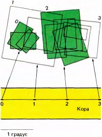
Rice. 92. When an electrode is moved in the cat’s cortex over a long distance parallel to the surface, the position of the receptive fields of the cells encountered changes. In this case, the electrode traveled 3 mm and encountered approximately 60 cells (too many to show in this figure). Here the positions of only four or five receptive fields are marked, maps for which were drawn at the beginning of the passage of each millimeter (about a tenth of it), and the remaining fields were left without attention. The upper part of the figure shows the receptive fields of cells encountered at four stages of passage (0, 1, 2 and 3). Each such group of fields noticeably shifted in the field of view to the right relative to the previous group. The receptive fields of cells in group 2 do not overlap with the fields of group 0, and the fields of group 3 do not overlap with the fields of group 1. In both cases, the distance along the cortex was 2 millimeters.
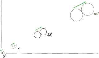
Rice. 93. In macaques, the receptive fields of cells in the upper layers of the cortex increase with distance from the fovea (which corresponds to 0°). The distance between the centers of the receptive fields of those cells, the distance between which in the cortex is 2 millimeters, also increases in the same ratio.
Functional units of the cortex
We have to conclude that any area of the primary visual cortex approximately 2-2 millimeters in size must have all the mechanisms necessary to fully process a certain part of the visual field, which is very small in the region of the fovea and much larger in the periphery. This region, which probably receives tens of thousands of input fibers from the lateral geniculate body, processes the information coming through them and sends output signals along fibers sensitive to the orientation of the stimulus, its movement, etc., combining information from both eyes. All sections perform similar operations on signals arriving via approximately the same number of fibers. The information received by a particular area is very detailed when it comes from a small area in the region of the fovea, and less detailed when it comes from a larger area in the periphery of the retina. The output signals of such a section are generated without any consideration of what size part of the field of view is analyzed here and with what degree of detail. The set of mechanisms for processing information in such areas is basically the same. This allows us to explain the homogeneity that we see both with the naked eye and during microscopic examination.
The fact that a shift along the cortex of 2 mm is just enough to enter the projection of a new area of the retina means that whatever local operations are performed in the cortex, they must all be performed within a block measuring 2-2 mm. Obviously, a smaller area will not be able to comprehensively analyze a correspondingly smaller area of the field of view, since such an analysis requires all the elements of the 2-mm block. This is clear already from considering the data on the position and size of the receptive fields. However, we will need to discuss in more detail what the words “analysis” and “operation” mean. We can start by looking at the line orientation parameter. In any part of the field of view, even the smallest, all possible orientations must be taken into account. If, when analyzing a certain area with a corresponding 2 mm cortical block, this block does not contain an element that responds to an orientation of +45°, then the presence of such an element in other cortical blocks will not help, since other blocks deal with other parts of the visual field. However, fortunately, the width of the orientation stripes in the cortex is quite small - 0.05 mm, and a set of such stripes for all orientations from 0 to 180° with a step of 10° is laid twice on a section of the cortex 2 mm wide. The situation is the same with ocular dominance - the width of the corresponding columns is 0.5 mm, so a 2 mm block is more than enough for a complete analysis. Thus, blocks with a diameter of 2 mm apparently have a complete set of necessary mechanisms.
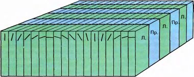
Rice. 94. We call this pattern “our ice cube model.” The diagram shows how the cortex is simultaneously divided into two types of plates - for ocular dominance (for the right and left eyes) and for orientation. This model should not be taken literally - none of these plate systems are all that regular, especially the orientation plates, which are far from being parallel and having straight boundaries. Moreover, they apparently do not intersect at any particular angle, and in any case do not form an orthogonal system like the one shown in the figure.
It should be added that blocks 2 mm in size are a feature not so much of the entire field 17 of the cortex, but of layer 3 of field 17. If we turn to layers 5 and 6, then the sizes of the receptive fields of the cells and their scatter are twice as large; therefore, for comprehensive analysis in layers 5 and 6 and the formation of receptive fields of larger magnitude and with more complex properties, blocks with a diameter of 4 mm will apparently be required. On the other hand, in layer 4C the receptive fields and their scatter are much smaller and, accordingly, the block sizes are closer to 0.1–0.2 mm. However, the general idea of individual functional units remains the same, regardless of the fact that any given area of the visual field is served by a set of local mechanisms located in several layers of the cortex, i.e. that the cortex is a collection of several systems combined into one whole.
All these considerations help us understand why the columns cannot be much larger. In a block of 2×2 mm in size it is necessary to place all the mechanisms that highlight the necessary variables in the stimuli. So far we've talked about orientation and ocular dominance, but in reality, more than two input variables are mapped onto a two-dimensional surface. For this purpose, two variables were selected as the main parameters that determine the coordinates in the field of view (distances from the central fovea horizontally and vertically). The resulting coordinate grid additionally displays other variables, such as stimulus orientation and ocular dominance, in the form of a finer mosaic.
We will call a section of cortex with a diameter of 2 mm module. Personally, this term does not seem entirely appropriate to me, in particular because it evokes an overly specific idea of a small rectangular block of an electronic device, which, together with hundreds of other similar blocks, is mounted on a common chassis. However, to a certain extent, we had in mind precisely this association, but of a more generalized nature. Firstly, the functional modules of the orientation system that we have identified are such that the boundaries between adjacent modules are conditional. For example, we can consider that a module begins with cells that highlight vertical orientation and ends with the next group of cells that also highlight vertical orientation; Any other orientation can be taken as a starting point, as long as all orientations are represented at least once in each module. The situation is the same with ocular dominance - the module can begin from the place where the right eye is dominant, from where the left eye is dominant, or from the middle of the column, as long as the module ultimately includes two sections of ocular dominance, one for each eye. Secondly, as we have already mentioned, the magnitude of the modulus will depend on which of the layers of the cortex we are considering. However, the term "module" should connote a system of about 500–1000 miniature devices, all of which are interchangeable (assuming that about 10,000 input and probably about 50,000 output connections could be reproduced).
It must be added right away that no one, of course, thinks that the cortex is completely homogeneous, from the projection of the central fovea to the areas representing the far periphery. In addition to visual acuity, other parameters of visual perception also change as you move away from the center of the retina; For example, color vision deteriorates, although perhaps not very sharply, if you compensate for the decrease in the “gain” coefficient (i.e., increase the size of objects as they move away from the point of fixation). Movement, as well as very faint light sources, are likely to be more easily noticed at the periphery of the visual field. Binocular vision should deteriorate, since in zones with a radius from 20 to 80° the ocular dominance columns for the ipsilateral eye become narrower and narrower, and for the contralateral eye - wider and wider. At distances greater than 80° from the fovea, the ipsilateral columns completely disappear and the cortical mechanisms become “monocular.” In view of these, as well as other differences that undoubtedly exist, the corresponding cortical structures must be somewhat heterogeneous. Therefore, cortical modules are probably not all exactly the same.
Cortical deformation
If you compare the cortex with the retina, you can get a more complete picture of the configuration of the cortex. The eye is a sphere because it is a purely optical instrument. Therefore, the shape of the retina is also spherical. In a camera, the film can be flat, since the lens aperture is on average about 30°. A wide-angle fisheye camera has a much larger aperture, but the image is distorted in the periphery. Of course, spherical photographs would be inconvenient - flat photographs are much more convenient to store. As for the eye, its spherical shape seems ideal, since the ball is a compact body, and at the same time it can easily rotate in the socket (if the eye had the shape of a cube, then difficulties would arise). And since the eye has a spherical shape, the angular scale of the image of the visual field on the retina remains constant everywhere - the number of degrees per 1 mm of the retina is the same on the entire retina. In humans it is 3.5°. As already mentioned, the centers of the receptive fields of ganglion cells in and near the fovea are very small, but they increase with distance from it. Therefore, we should not be surprised that in the central region of the retina there should be many more ganglion cells per millimeter than in the far periphery. Indeed, near the central fovea, these cells are laid out in several layers, while at the periphery of the retina they are located so rarely that visually they do not even form one continuous layer (see micrographs in Fig. 95). Since the retina must have a spherical shape, its layers cannot be uniform. This may be one reason that only very limited information processing occurs in the retina; otherwise, a very large thickening of its layers in the central region would be required.
In this sense, the cortex has greater capabilities. Unlike the retina, it may not be spherical, but may simply stretch in the area of the projection of the central fovea compared to the projection of the periphery. This stretching appears to be carried out to such an extent that the thickness of the cortical layer (as well as the width of the columns and other parameters) is the same everywhere.
What effect does this have on the overall shape of the cortex? Although I have repeatedly characterized the bark as a "layer", this does not mean that it must be flat. If there were no configuration distortions, the striate cortex would have a spherical shape - the same as that of the eyeball. In this sense, it is similar to the surface of the Earth - without the distortions associated with the relief, the surface of the Earth would have a strictly spherical shape. True, only about half of the posterior surface of the eye is reflected on the striate cortex of one hemisphere, i.e. approximately a quarter of the entire sphere.
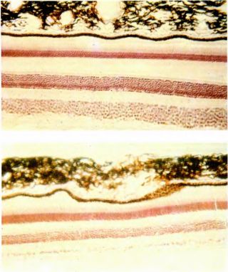
Rice. 95. Unlike the layers of the cortex, the layers of the retina are far from equal in thickness in its different parts. In both monkeys and humans, the layer of ganglion cells near the fovea (lower layer in the upper microphotograph) is 8–10 cells thick, while at the far periphery of the retina, at a distance of 70–80° from its center (lower microphotograph), ganglion cells do not form even one continuous layer. This should not surprise us, since the centers of the receptive fields of ganglion cells near the fovea are very small, and in the periphery they increase. In the center of the retina there are many more ganglion cells per unit area.
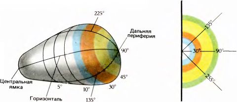
Rice. 96. The straightened striate cortex would be shaped like a pear. It would have the appearance of a quarter sphere if all parts of the retina were represented in it proportionally. In reality, however, the shape of the cortex is significantly distorted due to the disproportionately increased display of the central retinal region. The far periphery (approximately 90° from the center) is displayed so poorly that the semicircle corresponding to 90° (far right) appears very small.
By stretching so as to maintain constant thickness and yet be able to process the particularly abundant information from the many ganglion cells of the fovea, the cortex takes on a shape other than spherical. If you unfold and smooth out all the folds of the bark, its shape will be neither spherical nor flat. In reality, the cortex will then have the appearance of a highly distorted segment of a sphere, constituting a fourth of its part; in shape it will more likely resemble a pear or an egg. This result was predicted in 1962 by Daniel and Whitteridge, who experimentally determined the dependence of the “increase” in field 17 on the distance to the projection of the fovea (this was discussed on p. 0127). From the data obtained, they calculated the three-dimensional shape of the surface of the cortex, and then made a rubber model using a series of sequential histological sections. They then actually straightened out the model, thereby confirming their prediction of a pear-shaped cortex. The resulting form is shown in Fig. 96. Before this, no one had asked the question what shape the visual cortex would have if its folds were straightened. As far as I know, no one has also realized that any area of the cortex must have some kind of certain a form that logically follows from the functions of this area. Apparently, the folds of the cortex, which must be smoothed out (without stretching or tearing) to reveal the true shape, are formed because a large segment of the cortex had to be “crumpled” to fit into the limited space of the cranium. In this case, the nature of the folds may not be entirely random; some details are probably determined by the fact that intercortical connections should be as short as possible.
If we turn to the somatosensory cortex, here the complexity of topographic mapping can reach an extreme degree. For example, the area of the cortex corresponding to the skin of the hand should be similar in configuration to a glove, and with certain distortions in order to provide greater sensitivity to the fingertips compared to the palm and back of the hand. This distortion is analogous to an increase in the cortical projection of the fovea compared to the periphery of the retina in accordance with differences in visual acuity. Will the cortical projection of the hand really resemble a glove if we make a rubber model and gradually inflate it to straighten out the artificial folds? Probably not. Mapping the somatosensory cortex has proven to be extremely challenging. The results obtained so far suggest that the form of a continuous projection would be too bizarre; in fact, the somatosensory cortex seems to be cut with scissors into many small, more “convenient” sections, which are connected like pieces of a patchwork quilt so that some approximation of a flat surface is obtained.
Eye, brain, vision