Corpus callosum and stereoscopic vision
Corpus callosum is a powerful bundle of myelinated fibers connecting the two hemispheres of the brain. Stereoscopic vision (stereopsis) is the ability to perceive the depth of space and assess the distance of objects from the eyes. These two things are not particularly closely related, but it is known that a small part of the fibers of the corpus callosum does play some role in stereopsis. It turned out to be convenient to include both of these topics in one chapter, since when considering them we will have to take into account the same feature of the structure of the visual system, namely, that in the chiasm there are both crossed and uncrossed fibers of the optic nerve (see Fig. 35).
Corpus callosum
The corpus callosum (corpus callosum in Latin) is the largest bundle of nerve fibers in the entire nervous system. According to a rough estimate, there are about 200 million axons in it. The true number of fibers is likely even higher, since the estimate is based on conventional light microscopy rather than electron microscopy. This number is incomparable to the number of fibers in each optic nerve (1.5 million) and in the auditory nerve (32,000). The cross-sectional area of the corpus callosum is about 700 mm2, whereas in the optic nerve it does not exceed a few square millimeters. The corpus callosum, together with a thin bundle of fibers called anterior commissure, connects the two hemispheres of the brain (Fig. 98 and 99). Term commissary means a set of fibers connecting two homologous nerve structures located in the left and right halves of the brain or spinal cord. The corpus callosum is also sometimes called the greater commissure of the brain.
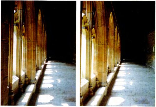
Rice. 97. This stereo pair shows a gallery at New College, Oxford. After this was done, we moved it 7.5 cm to the left and took the left photo.
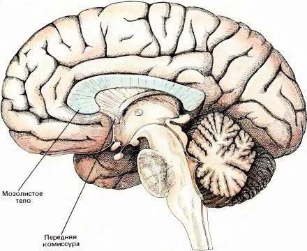
Rice. 98. The human brain in a section along the median plane. A thick, arched in cross-section bundle of axons is visible - the corpus callosum.
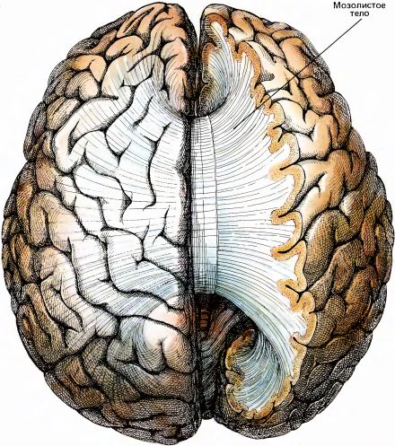
Rice. 99. Here the brain is shown from above. Part of the right hemisphere is cut away, and a bundle of fibers of the corpus callosum is visible, connecting all parts of the two hemispheres.
Until about 1950, the role of the corpus callosum was completely unknown. In rare cases, there is a congenital absence (aplasia) corpus callosum. This formation can also be partially or completely cut during a neurosurgical operation, which is done deliberately - in some cases in the treatment of epilepsy (so that a convulsive discharge occurring in one hemisphere of the brain cannot spread to the other hemisphere), in other cases in order to reach a deep-lying tumor from above (if, for example, the tumor is located in the pituitary gland). According to the observations of neurologists and psychiatrists, after this type of operation no mental disorders occur. Some have even suggested (though hardly seriously) that the sole function of the corpus callosum is to hold the two hemispheres of the brain together. Until the 1950s, little was known about the details of the distribution of connections in the corpus callosum. It was obvious that the corpus callosum connects the two hemispheres, and on the basis of data obtained by rather crude neurophysiological methods, it was believed that in the striate cortex the fibers of the corpus callosum connect exactly symmetrical areas of the two hemispheres.
In 1955, Ronald Myers, a graduate student of psychologist Roger Sperry at the University of Chicago, pioneered an experiment that revealed some of the functions of this huge fiber tract. Myers trained cats by placing them in a box with two side-by-side screens on which different images could be projected, such as a circle on one screen and a square on the other. The cat was trained to put its nose on the screen that showed a circle and ignore the other screen that showed a square. Correct answers were reinforced with food, and for incorrect answers the cats were slightly punished - a loud bell was turned on, and the cat was not rudely, but decisively pulled away from the screen. With this method, over several thousand repetitions, the cat can be brought to the level of reliable discrimination of figures. (Cats learn slowly; for example, pigeons need from several tens to several hundred repetitions to learn a similar task, but a person can generally be taught immediately by giving him verbal instructions. This difference seems somewhat strange - after all, a cat has a brain many times larger than a pigeon.)
It is not surprising that Myers's cats learned to solve this problem just as well when one of the animal's eyes was covered with a mask. It is also not surprising that if training in such a task as choosing a triangle or a square was carried out with only one eye open - the left one, and during testing the left eye was closed and the right one was opened, then the accuracy of discrimination remained the same. This does not surprise us because we ourselves can easily solve a similar problem. The ease of solving such problems is understandable if we take into account the anatomy of the visual system. Each hemisphere receives input from both eyes. As we already said in Chapter 4, most of the cells in field 17 also have inputs from both eyes. Myers created a more interesting situation by performing a longitudinal section of the chiasm along the midline. Thus, he cut the intersecting fibers and kept the non-intersecting ones intact (this operation requires a certain skill from the surgeon). As a result of such a transection, the animal’s left eye was connected only to the left hemisphere, and the right eye only to the right. The idea of the experiment was to train the cat using the left eye, and in the “exam”, address the stimulus to the right eye. If the cat can solve the problem correctly, this will mean that the necessary information is transmitted from the left hemisphere to the right along the only known path - through the corpus callosum. So Myers cut the chiasm longitudinally, trained the cat with one eye open, and then tested it by opening the other eye and closing the first. Under these conditions, the cats still successfully solved the problem. Finally, Myers repeated the experiment on animals in which both the chiasm and the corpus callosum had previously been cut. This time the cats did not solve the problem. Thus, Myers experimentally established that the corpus callosum did perform some functions (although one could hardly think that it existed only so that individuals or animals with a severed optic chiasm could solve certain problems using one eye after learning using the other).
Study of the physiology of the corpus callosum
One of the first neurophysiological studies in this area was carried out several years after Myers' experiments by D. Whitteridge, then working in Edinburgh. Whitteridge reasoned that there was little reason for bundles of nerve fibers to connect homologous mirror-symmetrical sections of fields 17. Indeed, there is no reason for a nerve cell in the left hemisphere, connected to some points in the right half of the visual field, to connect with a cell in the right hemisphere, connected to a symmetrical section of the left half of the visual field. To test his assumptions, Whitteridge cut the optic tract on the right side of the brain behind the chiasm, thereby blocking the path of input signals to the right occipital lobe; but this, of course, did not exclude the transmission of signals there from the left occipital lobe through the corpus callosum (Fig. 100). Whitteridge then began to turn on the light stimulus and use a metal electrode to record electrical activity from the surface of the cortex. He actually received answers in his experience, but they appeared only on the inner border of field 17, i.e. in an area receiving input from a long, narrow vertical stripe in the middle of the visual field: when stimulated with small spots of light, responses appeared only when the light flashed on or near the vertical midline. If the cortex of the opposite hemisphere was cooled, thereby temporarily suppressing its function, the responses stopped; This was also caused by cooling of the corpus callosum. Then it became clear that the corpus callosum cannot connect the entire field 17 of the left hemisphere with the entire field 17 of the right hemisphere, but connects only small areas of these fields, where the projections of the vertical line are located in the middle of the visual field.
A similar result could have been predicted based on a number of anatomical data. Only one portion of area 17, located very close to the border with area 18, sends axons through the corpus callosum to the other hemisphere, and most of them seem to terminate in area 18 near the border with area 17. If we assume that the inputs to the cortex from the NKT exactly correspond to the contralateral parts of the visual field (namely, the left hemifield is mapped to the cortex of the right hemisphere, and the right hemifield to the cortex of the left hemisphere), then the presence connections between the hemispheres through the corpus callosum should ultimately lead to the fact that each hemisphere will receive signals from an area slightly larger than half the visual field. In other words, due to connections through the corpus callosum, there will be an overlap of hemifields projected into the two hemispheres. This is exactly what we found. Using two electrodes inserted into the cortex at the border of fields 17 and 18 in each hemisphere, we were often able to record the activity of cells whose receptive fields overlapped by several angular degrees.
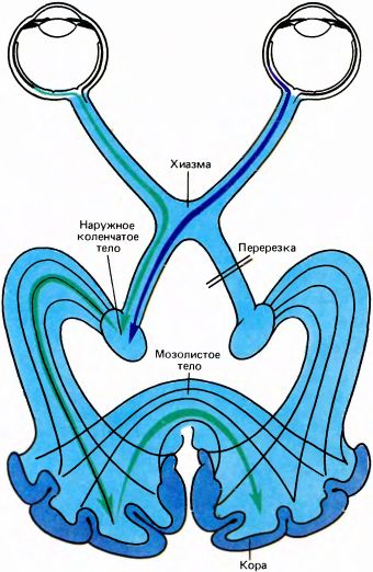
Rice. 100. In his experiment, Whitteridge performed a transection of the right optic tract. Now, in order for information to enter the visual cortex of the right hemisphere, it must first enter the visual cortex of the left hemisphere, and then pass to the other hemisphere through the corpus callosum. Cooling any part of this pathway blocks the transmission of nerve impulses.
T. Wiesel and I soon made microelectrode leads directly from the area of the corpus callosum (in its very posterior part) where there are fibers associated with the visual system. We found that almost all the fibers that we could activate with visual stimuli responded exactly like normal area 17 neurons, i.e. exhibited the properties of both simple and complex cells, selectively sensitive to stimulus orientation and usually responding to stimulation of both eyes. In all these cases, the receptive fields were located very close to the mid-vertical below or above (or at the level of) the fixation point, as shown in Fig. 101.
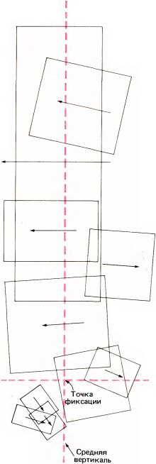
Rice. 101. The receptive fields of the fibers of the corpus callosum lie very close to the middle vertical of the visual field. All of these fields were identified by recording the activity of ten fibers in one cat.
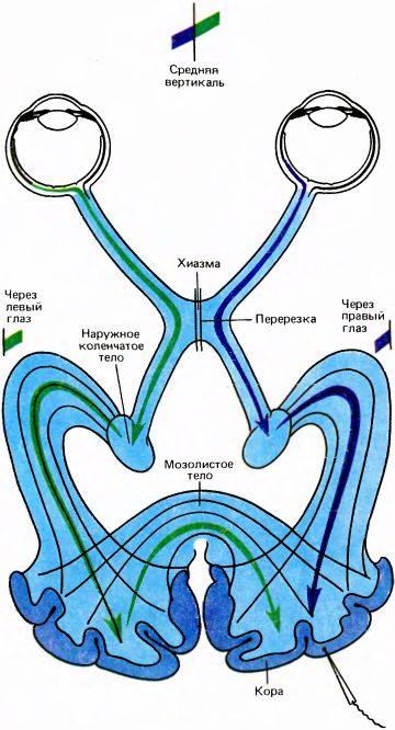
Rice. 102. Berlucchi and Rizzolatti in their experiment elegantly demonstrated not only the function of the visual part of the fibers of the corpus callosum, but also the high specificity of interhemispheric connections between cells similar in orientation and location of receptive fields. Berlucchi and Rizzolatti cut the cat's chiasm along the midline, so that the left eye began to transmit information only to the left hemisphere, and this information concerned only the right half of the visual field. Likewise, the right eye began to provide information only to the right hemisphere, and this information applied only to the left half of the visual field. After transection, responses were recorded from those cells whose receptive fields under normal conditions would intersect the middle vertical. It turned out that the receptive fields of such cells are split along this vertical; the right side is now connected to the left eye, and the left to the right eye.
Perhaps the most elegant neurophysiological demonstration of the role of the corpus callosum was the work of G. Berlucchi and G. Rizzolatti from Pisa, performed in 1968. Having cut the optic chiasm along the midline, they recorded responses in area 17 near the border with area 18, looking for those cells that could be activated binocularly. It is clear that any binocular cell in this area in the right hemisphere must receive input signals both directly from the right eye (via the NKT) and from the left eye and left hemisphere through the corpus callosum. As it turned out, the receptive field of each binocular cell captured the middle vertical of the retina, and that part of it that belongs to the left half of the visual field delivered information from the right eye, and that part that goes into the right half - from the left eye. Other properties of the cells studied in this experiment, including orientation selectivity, turned out to be identical (Fig. 102).
The results clearly showed that the corpus callosum connects cells to each other in such a way that their receptive fields can extend both to the right and to the left of the middle vertical. Thus, it seems to glue two halves of the image of the surrounding world. To better imagine this, let us assume that initially the cortex of our brain formed as one whole, not divided into two hemispheres. In this case, field 17 would have the appearance of one continuous layer onto which the entire visual field would be mapped. Then neighboring cells, in order to realize such properties as, for example, sensitivity to movement and orientation selectivity, would, of course, have to have a complex system of mutual connections. Now let’s imagine that the “designer” (be it God, or, say, natural selection) decided that it cannot be left like this any longer - from now on, half of all cells should form one hemisphere, and the other half - the other hemisphere. What then must be done with all the multitude of intercellular connections if two sets of cells must now move away from each other? Apparently, you can simply stretch these connections, forming part of the corpus callosum from them. In order to eliminate the delay in transmitting signals along such a long path (about 12-15 centimeters in humans), it is necessary to increase the transmission speed by providing the fibers with a myelin sheath. Of course, nothing of this sort actually happened during evolution; long before the cortex arose, the brain already had two separate hemispheres.
The experiment of Berlucchi and Rizzolatti, in my opinion, provided one of the most striking confirmations of the amazing specificity of neural connections. The cell shown in Fig. 108 (near the tip of the electrode) and probably a million other similar cells connected through the corpus callosum acquire their orientation selectivity both due to local connections with neighboring cells and due to connections going through the corpus callosum from the other hemisphere from cells with the same orientation sensitivity and similar arrangement of receptive fields (the above also applies to other properties of cells, such as directional specificity, the ability to respond to line ends, as well as complexity). Each of the cells in the visual cortex that have connections through the corpus callosum must receive input signals from cells in the other hemisphere with exactly the same properties. We know many facts indicating the selectivity of compounds in the nervous system, but I think that this example is the most striking and convincing.
The axons of the visual cortex cells discussed above constitute only a small proportion of all fibers of the corpus callosum. Experiments using axonal transport were carried out on the somatosensory cortex, similar to the experiments described in previous chapters with the injection of a radioactive amino acid into the eye. Their results indicate that the corpus callosum similarly connects those areas of the cortex that are activated by cutaneous and joint receptors located near the midline of the body on the trunk and head, but does not connect cortical projections of the limbs.
Each cortical area connects to several or even many other cortical areas of the same hemisphere. For example, the primary visual cortex is connected to area 18 (visual area 2), medial temporal area (area MT), visual area 4, and one or two other areas. Many areas of the cortex also have connections with several areas of the other hemisphere, through the corpus callosum, and in some cases through the anterior commissure. Therefore, we can consider these commissural connections simply as a special type of cortico-cortical connections. It is easy to imagine that this is demonstrated by such a simple example: if I tell you that my left hand feels cold or that I saw something to the left, then I formulate words using my cortical speech areas located in the left hemisphere (said, May be, and not entirely true, since I'm left-handed); information coming from the left half of the visual field or from the left hand is transmitted to my right hemisphere; then the corresponding signals must be transmitted through the corpus callosum to the speech zone of the cortex of the other hemisphere so that I can say something about my sensations. In a series of studies that began in the early 1960s, R. Sperry (now at the California Institute of Technology) and his colleagues showed that a person whose corpus callosum is cut (to treat epilepsy) loses the ability to talk about events about which information enters the right hemisphere. Work with such subjects has become a valuable source of new information about various functions of the cortex, including thinking and consciousness. The first articles about this appeared in the magazine Brain; they are extremely interesting, and can be easily understood by anyone who has read the real book.
Stereoscopic vision
The distance estimation mechanism, based on the comparison of two retinal images, is so reliable that many people (unless they are psychologists or specialists in visual physiology) are not even aware of its existence. To see the importance of this mechanism, try driving a car or bicycle, playing tennis or skiing for a few minutes with one eye closed. Stereoscopes have fallen out of fashion and you can only find them in antique stores. However, most readers watched stereoscopic films (when the viewer has to wear special glasses). The operating principle of both a stereoscope and stereoscopic glasses is based on the use of the stereopsis mechanism.
Images on the retinas are two-dimensional, but we see the world in three dimensions. Obviously, the ability to determine the distance to objects is important for both humans and animals. Similarly, perceiving the three-dimensional shape of objects means judging relative depth. Let's take a round object as a simple example. If it is located obliquely relative to the line of sight, its image on the retinas will be elliptical, but usually we easily perceive such an object as round. This requires the ability to perceive depth.
Humans have many mechanisms for judging depth. Some of them are so obvious that they hardly deserve mention. Nevertheless, I will mention them. If the size of an object is approximately known, for example in the case of objects such as a person, a tree or a cat, then we can estimate the distance to it (although there is a risk of error if we encounter a dwarf, a dwarf tree or a lion). If one object is located in front of another and partially obscures it, then we perceive the front object as being closer. If you take a projection of parallel lines, for example, railway rails, going into the distance, then in the projection they will come closer. This is an example of perspective, a very effective indicator of depth. A convex section of a wall appears lighter in its upper part if the light source is located higher (usually light sources are located at the top), and a recess in its surface, if illuminated from above, appears darker in the upper part. If the light source is placed at the bottom, then the convexity will look like a recess, and the recess will look like a convexity. An important sign of remoteness is motion parallax - the apparent relative displacement of near and more distant objects if the observer moves his head left and right or up and down. If any solid object is rotated, even at a small angle, its three-dimensional shape is immediately revealed. If we focus the lens of our eye on a nearby object, then a more distant object will be out of focus; thus changing the shape of the lens, i.e. By changing the accommodation of the eye (see Chapters 2 and 6), we get the opportunity to assess the distance of objects. If you change the relative direction of the axes of both eyes, bringing them together or spreading them apart (carrying out convergence or divergence), then you can bring together two images of an object and hold them in this position. Thus, by controlling either the lens or the position of the eyes, it is possible to estimate the distance of an object. The designs of a number of rangefinders are based on these principles. With the exception of convergence and divergence, all other distance measures listed so far are monocular. The most important mechanism of depth perception, stereopsis, depends on the joint use of the two eyes. When viewing any three-dimensional scene, the two eyes form slightly different images on the retina. You can easily verify this if you look straight ahead and quickly move your head from side to side by about 10 cm, or quickly close one eye or the other. If you have a flat object in front of you, you won't notice much of a difference. However, if the scene includes objects at different distances from you, you will notice significant changes in the picture. During stereopsis, the brain compares images of the same scene on two retinas and estimates relative depth with great accuracy.
Suppose the observer fixes with his gaze a certain point P. This statement is equivalent to if we say: the eyes are directed in such a way that the images of the point appear in the central fossa of both eyes (F in Fig. 103). Suppose now that Q is another point in space that appears to the observer to be located at the same depth as P. Let QL and QR — images of the Q point on the retinas of the left and right eyes. In this case, points QL and QR called corresponding points two retinas. Obviously, two points coinciding with the central fovea of the retina will be corresponding. From geometric considerations it is also clear that the point Q', assessed by the observer as located closer than Q, will give two projections on the retinas - Q'L and Q'R - at non-corresponding points located further from each other than if these points were corresponding (this situation is shown on the right side of the figure). In the same way, if we consider a point located further from the observer, it turns out that its projections on the retinas will be located closer to each other than the corresponding points. What is said above about the corresponding points is partly definitions, and partly statements arising from geometric considerations. When considering this issue, the psychophysiology of perception is also taken into account, since the observer subjectively assesses whether the object is located further or closer to point P. Let's introduce one more definition. All points that, like point Q (and, of course, point P), are perceived as equidistant, lie on horoptera - a surface passing through points P and Q, the shape of which differs from both a plane and a sphere and depends on our ability to estimate distance, i.e. from our brain. Distances from the fovea F to the projections of point Q (QL and QR) are close, but not equal. If they were always equal, then the line of intersection of the horopter with the horizontal plane would be a circle.
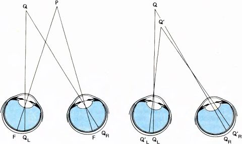
Rice. 103. Left: if the observer looks at point P, then two of its images (projections) fall on the central fossa of the two eyes (point F). Q is a point that, according to the observer, is at the same distance from him as P. In this case, they say that two projections of the point Q (QL and QR) fall into the corresponding points of the retinas. (The surface composed of all points Q that appear to be at the same distance from the observer as point P is called a horopter passing through point P). Right: if point Q' is closer to the observer than Q, then its projections on the retinas (Q'L and Q'R) will be further apart horizontally than if they were at corresponding points. If point Q' were further away, then the projections Q'L and Q'R would be shifted horizontally closer to each other.
Let us now assume that we fix with our gaze a certain point in space and that in this space there are two point sources of light, which give a projection on each retina in the form of a point of light, and these points are not corresponding: the distance between them is several more, than between corresponding points. We will call any such deviation from the position of the corresponding points disparity. If this deviation in the horizontal direction does not exceed 2° (0.6 mm on the retina), and in the vertical direction no more than several arc minutes, then we will visually perceive a single point in space located closer than the one we are fixing. If the distances between the projections of the point are no greater, but less, than between corresponding points, then this point will seem located further than the fixation point. Finally, if the vertical deviation exceeds several arc minutes or the horizontal deviation is more than 2°, then we will see two separate points that may seem to be located further or closer to the fixation point. These experimental results illustrate the basic principle of stereo perception first formulated in 1838 by Sir C. Wheatstone (who also invented the device known in electrical engineering as the “Wheatstone bridge”).
It seems almost incredible that, until this discovery, no one seemed to realize that the presence of subtle differences in the images projected on the retinas of the two eyes could give rise to a distinct impression of depth. Such a stereo effect can be demonstrated in a few minutes by anyone who can arbitrarily move the axes of their eyes together or apart, or by someone who has a pencil, a piece of paper and several small mirrors or prisms. It is unclear how Euclid, Archimedes and Newton missed this discovery. In his article, Wheatstone notes that Leonardo da Vinci was very close to discovering this principle. Leonardo pointed out that a ball located in front of any spatial scene is seen differently by each eye - with the left eye we see its left side a little further, and with the right eye we see the right side. Wheatstone further notes that if Leonardo had chosen a cube instead of a ball, he would certainly have noticed that its projections were different for different eyes. After this, he might, like Wheatstone, become interested in what would happen if two similar images were specially projected onto the retinas of two eyes.
An important physiological fact is that the sensation of depth (i.e., the ability to “directly” see whether an object is further or closer to the point of fixation) occurs in cases where two retinal images are slightly displaced relative to each other in the horizontal direction - moved apart or, conversely, brought together (unless this displacement exceeds about 2 °, and the vertical displacement is close to zero). This, of course, corresponds to geometric relationships: if, relative to a certain distance reference point, an object is located closer or further, then its projections on the retinas will be moved apart or brought closer together horizontally, while no significant vertical displacement of the images will occur.
This is the basis of the action of the stereoscope invented by Wheatstone. The stereoscope was so popular for about half a century that it was found in almost every home. The same principle underlies the stereo cinema that we now watch using special Polaroid glasses. In the original design of the stereoscope, the observer viewed two images placed in a box using two mirrors that were positioned so that each eye saw only one image. For convenience, prisms and focusing lenses are now often used. The two images are identical in every way except for slight horizontal offsets, which create the impression of depth. Anyone can produce a photograph suitable for use in a stereoscope by selecting a stationary object (or scene), taking a photograph, and then moving the camera 5 centimeters to the right or left and taking a second photograph.
Not everyone has the ability to perceive depth using a stereoscope. You can easily check your stereopsis yourself if you use the stereo pairs shown in Fig. 105 and 106. If you have a stereoscope, you can make copies of the stereo pairs shown here and paste them into the stereoscope. You can also place a thin piece of cardboard perpendicularly between two images from the same stereo pair and try to look at your image with each eye, setting your eyes parallel, as if you were looking into the distance. You can also learn to move your eyes together and apart using your finger, placing it between your eyes and the stereo pair and moving it forward or back until the images merge, after which (this is the most difficult) you can examine the merged image, trying not to split it into two. If you can do this, the apparent depth relationships will be the opposite of those perceived when using a stereoscope.
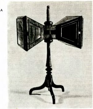
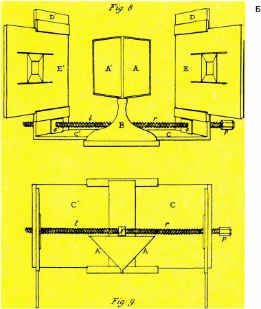
Rice. 104. A. Wheatstone stereoscope. B. Diagram of Wheatstone's stereoscope, compiled by himself. The observer sits in front of two mirrors (A and A'), placed at an angle of 40° to the direction of his gaze, and looks at two pictures combined in the field of view - E (with the right eye) and E' (with the left eye). In a simpler version created later, two pictures are placed side by side so that the distance between their centers is approximately equal to the distance between the eyes. The two prisms deflect the direction of gaze so that, with proper convergence, the left eye sees the left image and the right eye sees the right image. You yourself can try to do without a stereoscope, imagining that you are looking at a very distant object with eyes whose axes are set parallel to each other. Then the left eye will look at the left image, and the right eye will look at the right one.
Even if you can't replicate the depth perception experiment, either because you don't have a stereoscope or because you can't voluntarily move the axes of your eyes in and out, you can still get the idea, although you won't enjoy the stereo effect.
In the upper stereo pair in Fig. 105 in two square frames there is a small circle, one of which is shifted slightly to the left of the center, and the other slightly to the right. If you examine this stereopair with both eyes, using a stereoscope or another method of combining images, you will see a circle not in the plane of the sheet, but in front of it at a distance of about 2.5 cm. If you also examine the lower stereopair in Fig. 105, then the circle will be visible behind the plane of the sheet. You perceive the position of the circle in this way because the retinas of your eyes receive exactly the same information as if the circle really was in front or behind the plane of the frame.
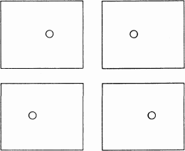
Rice. 105. If the upper stereo pair is inserted into a stereoscope, the circle will appear to be located in front of the plane of the frame. In the lower stereo pair it will be located behind the plane of the frame. (You can do this experiment without a stereoscope, by convergence or divergence of the eyes; for most people, convergence is easier. To make the task easier, you can take a piece of cardboard and place it between two images of a stereo pair. At first, this exercise may seem difficult and tedious to you; do not try too hard the first time. When converging the eyes, the circle on the upper stereo pair will be visible further than the plane, and on the lower one, closer).
In 1960, Bela Jules of Bell Telephone Laboratories came up with a very useful and elegant technique for demonstrating the stereo effect. The image shown in Fig. 107, at first glance appears to be a homogeneous random mosaic of small triangles. This is true, except that there is a larger hidden triangle in the central part. If you view this image with two pieces of colored cellophane placed in front of your eyes - red in front of one eye and green in front of the other, then you should see a triangle in the center protruding forward from the plane of the sheet, as in the previous case with the small circle on the stereo pairs. (You may have to watch for a minute or so the first time until the stereo effect occurs.) If you swap the pieces of cellophane, a depth inversion will occur. The value of these Yulesz stereo pairs is that if you have impaired stereo perception, you will not see the triangle in front of or behind the surrounding background.
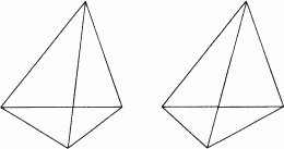
Rice. 106. Another stereo pair.
To summarize, we can say that our ability to perceive the stereo effect depends on five conditions:
1. There are many indirect signs of depth - partial obscuring of some objects by others, motion parallax, rotation of an object, relative sizes, casting shadows, perspective. However, the most powerful mechanism is stereopsis.
2. If we fix our gaze on some point in space, then the projections of this point fall into the central fossa of both retinas. Any point that is judged to be located at the same distance from the eyes as the point of fixation forms two projections at corresponding points on the retinas.
3. The stereo effect is determined by a simple geometric fact - if some object is closer to the point of fixation, then its two projections on the retinas are farther from each other than the corresponding points.
4. The main conclusion, based on the results of experiments with subjects, is the following: an object whose projections on the retinas of the right and left eyes fall on the corresponding points is perceived as located at the same distance from the eyes as the fixation point; if the projections of this object are moved apart compared to the corresponding points, the object appears to be located closer to the fixation point; if, on the contrary, they are close, the object appears to be located further than the point of fixation.
5. When the horizontal displacement of projections is more than 2° or the vertical displacement is more than several arc minutes, double vision occurs.
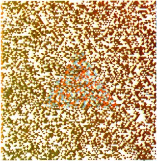
Rice. 107. In order to obtain this image, called anaglyph, Bela Jules first constructed two systems of randomly placed small triangles; they differed only in that 1) one system had red triangles on a white background, and the other had green triangles on a white background; 2) within a large triangular zone (near the center of the picture), all green triangles are slightly shifted to the left compared to red ones. After this, the two systems are combined, but with a slight shift, so that the triangles themselves do not overlap each other. If the resulting image is viewed through a green cellophane filter, only red elements will be visible, and if through a red filter, only green elements will be visible. If you place a green filter in front of one eye and a red filter in front of the other, you will see a large triangle protruding about 1 cm in front of the page. If the filters are swapped, the triangle will be visible behind the page plane.
Physiology of stereoscopic vision
If we want to know what the brain mechanisms of stereopsis are, the easiest place to start is by asking: Are there neurons whose responses are specifically determined by the relative horizontal displacement of the images on the retinas of the two eyes? Let's first look at how the cells of the lower levels of the visual system respond when both eyes are simultaneously stimulated. We must start with neurons in area 17 or higher because retinal ganglion cells are clearly monocular, and cells in the lateral geniculate body, in which input from the right and left eyes are distributed in different layers, can also be considered monocular—they respond to stimulation of either one eye or the other, but not both. In area 17, approximately half of the neurons are binocular cells that respond to stimulation of both eyes. Upon careful testing, it turns out that the responses of these cells seem to depend little on the relative position of the stimulus projections on the retinas of the two eyes. Consider a typical complex cell that responds with a continuous discharge to the movement of a stimulus strip through its receptive field in one eye or the other. When both eyes are simultaneously stimulated, the frequency of discharges of this cell is higher than when one eye is stimulated, but it is usually not important for the response of such a cell whether at any moment the stimulus projections fall into exactly the same parts of the two receptive fields. The best response is recorded when these projections enter and exit the respective receptive fields of the two eyes at approximately the same time; however, it is not so important which projection is slightly ahead of the other. In Fig. 108 shows a characteristic curve of the response (for example, the total number of impulses in the response during one passage of the stimulus through the receptive field) on the difference in the position of the stimulus on both retinas. This curve is very close to a horizontal straight line, which makes it clear that the relative position of the stimuli on the two retinas is not very significant. A cell of this type will respond well to a line of proper orientation regardless of its distance - the distance to the line may be greater than, equal to, or less than the distance to the point fixed by the gaze.
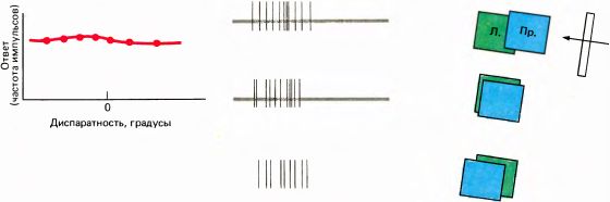
Rice. 108. When both eyes are simultaneously stimulated by a vertical light line moving to the left, an ordinary binocular cell in field 17 will give the same responses at three different relative positions of this line on both retinas. Zero disparity means that there is no difference in position—as if the monkey were looking at a screen on which the stimuli are presented. The absence of disparity does not lead to a noticeable increase in the cell response.
Compared to this cell, the neurons whose responses are presented in Fig. 109 and 110 are very sensitive to the relative position of the two stimuli on the two retinas, i.e. sensitive to depth. The first neuron (Fig. 109) responds best if the stimuli fall exactly on the corresponding areas of the two retinas. The amount of horizontal misalignment of stimuli (i.e., disparity) at which the cell stops responding is a certain fraction of the width of its receptive field. Therefore, the cell responds if and only if the object is approximately the same distance from the eyes as the fixation point. The second neuron (Fig. 110) responds only when the object is located further than the fixation point. There are also cells that respond only when the stimulus is located closer to this point. When the degree of disparity changes, neurons of the last two types, called distant cells и nearby cells very sharply change the intensity of their responses at or near the point of zero disparity. Neurons of all three types (cells, tuned to disparity) were discovered in field 17 monkeys. It is not yet entirely clear how often they occur there, whether they are located in certain layers of the cortex, and whether they are in certain spatial relationships to the ocular dominance columns. These cells are highly sensitive to the distance of an object from the eyes, which is encoded as the relative position of the corresponding stimuli on the two retinas. Another feature of these cells is that they do not respond to stimulation of only one eye or respond, but very weakly. All these cells have the common property of orientation selectivity; as far as we know, they are similar to ordinary complex cells of the upper layers of the cortex, but they have an additional property - sensitivity to depth. In addition, these cells respond well to moving stimuli and sometimes to the ends of lines.
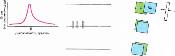
Rice. 109. There is a significant difference in the response of this selective responding cell depending on whether the stimulus is exactly the same distance as the fixation point, or whether it is closer or further away. This cell responds only when the distance to the stimulus is the same as to the fixation point. In this experiment, the axis of one eye was deflected horizontally using a prism; however, moving the screen toward or away from the animal would have the same effect.
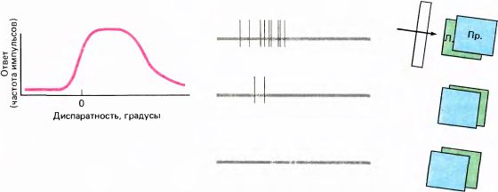
Rice. 110. In the type of cell presented here (“far”), objects located closer to the screen plane cause a very weak response or do not cause it at all. At zero disparity (i.e., when the distance to the stimulus is equal to the distance to the screen), a small displacement of the screen has a strong effect on the response: it increases sharply and remains constant for stimuli located further than the fixation point. If the stimulus is moved far enough away, then the two receptive fields of the cell no longer overlap, i.e., essentially, the two eyes are stimulated independently. In this case the response stops.
J. Poggio of Johns Hopkins Medical School recorded the responses of such cells in field 17 of an awake monkey with implanted electrodes, which had previously been trained to fixate a specific object with its gaze. In anesthetized monkeys, such cells were also detected in the cortex, but they were rarely found in area 17 and very often in area 18. I would be extremely surprised if it turned out that animals and humans can stereoscopically estimate distances to objects using only the three types of cells described above - tuned to zero disparity, “near” and “far”. I would rather expect to find a complete set of cells for all possible depths. In awake monkeys, Poggio also encountered narrowly tuned cells that responded best not to zero disparity, but to small deviations from it; Apparently, there may be specific neurons in the cortex for all levels of disparity. Although we still don't know exactly how the brain "reconstructs" a scene involving many widely spaced objects (whatever we mean by "reconstruction"), cells like those described above are likely involved in the early stages of this process.
Some problems associated with stereoscopic vision
During the study of stereopsis, psychophysicists encountered a number of problems. It turned out that the processing of some binocular stimuli occurs in the visual system in completely unclear ways. I could give many examples of this kind, but I will limit myself to just two.
Using the example of stereo pairs shown in Fig. 105, we saw that moving two identical images (in this case circles) towards each other leads to a feeling of greater proximity, and towards each other - to a feeling of greater distance. Let us now assume that we perform both of these operations simultaneously, for which we place two circles in each frame, located next to each other (Fig. 111). Obviously, considering such a stereopair could lead to the perception of two circles - one closer and the other further than the plane of fixation. However, we can assume another option: we will simply see two circles lying side by side in the plane of fixation. The fact is that these two spatial situations correspond to the same images on the retinas. In reality, this pair of stimuli can be perceived only like two circles in the plane of fixation, which is easy to verify if you achieve the merger of the square frames in Fig. by any means. 111. In exactly the same way, we can imagine a situation where we consider two chains of signs ?, say, six characters in a chain. If you look at them through a stereoscope, then in principle you can perceive any of a number of possible configurations depending on which sign? from the left chain will merge with a certain sign ? in the right chain. In fact, if we examine such a stereopair through a stereoscope (or in another way that creates a stereo effect), we will always see six signs ? in the plane of fixation. We still don't know how the brain resolves this ambiguity and chooses the simplest possible combination. Because of this kind of ambiguity, it is difficult to even imagine how we manage to perceive a three-dimensional scene that includes many branches of different sizes located at different distances from us. True, physiological evidence suggests that the task may not be so difficult, since different branches are likely to have different orientations, and we already know that cells involved in stereopsis are always orientation-selective.
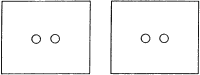
Rice. 111. Stereopairs shown in Fig. 105, caused the feeling that the circle was located closer or further than the plane of the frame. Here these two stereo pairs are combined, and therefore, it would seem that we should see one circle closer and the other further away. However, in reality this does not work; both circles are visible at the same distance as the frame.
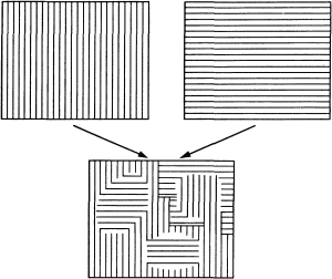
Rice. 112. This stereopair cannot be merged into a single perception, like other stereopairs (for example, those shown in Fig. 105 and 106). Instead, you get the effect of “struggle of visual fields” - you see a mosaic picture consisting of fragments of both images and constantly changing.
A second example of the unpredictability of binocular effects related to stereopsis is the so-called struggle of visual fields, which we also mention in the section on strabismus (chapter 9). If very different images are created on the retinas of the right and left eyes, then often one of them ceases to be perceived. If you look with your left eye at a grid of vertical lines and with your right eye at a grid of horizontal lines (Fig. 112; you can use a stereoscope or eye convergence), you would expect to see a grid of intersecting lines. However, in reality it is almost impossible to see both sets of lines at the same time. Either one or the other is visible, each of them only for a few seconds, after which it disappears and the other appears. Sometimes you can also see a mosaic of these two images, in which individual homogeneous areas will move, merge or separate, and the orientation of the lines in them will change (see Fig. 112, below). For some reason, the nervous system cannot perceive so many different stimuli simultaneously in the same area of the visual field, and it suppresses the processing of one of them. We use the word "suppress" here simply as another description of the same phenomenon: in fact, we do not know how such suppression occurs and at what level of the central nervous system it occurs. I think the mosaic nature of the perceived image as the visual fields compete suggests that the "decision making" in this process occurs quite early in the processing of visual information, perhaps in field 17 or 18. (I'm glad I don't have to defend this assumption.)
The phenomenon of visual field competition means that in cases where the visual system cannot combine the images on the two retinas (into a flat picture if the images are the same, or into a three-dimensional scene if there is only a slight horizontal disparity), it simply rejects one of the images - either completely, for example, when we look through a microscope with the other eye open, or partially or temporarily, as in the example described above. In the microscope situation, attention plays a significant role, but the neural mechanisms underlying this shift in attention are also unknown.
You can observe another example of the struggle between visual fields if you simply look at some multicolor scene or picture through glasses with red and green filters. The impressions of different observers in this case can be very different, but most people (including myself) note transitions from a general reddish tone to a greenish tone and back, but without the yellow color that is obtained when red light is usually mixed with green (see Chapter 8, Fig. 121).
Stereo blindness
If a person is blind in one eye, then it is obvious that he will not have stereoscopic vision. However, it is also absent in some people whose vision is otherwise normal. The surprising thing is that the proportion of such people is not too small. So, if you show stereo pairs like those shown in Fig. 105 and 106, with one hundred student subjects (using Polaroids and polarized light), it is usually found that four or five of them cannot achieve the stereo effect. This often surprises them, since in everyday conditions they do not experience any inconvenience. The latter may seem strange to anyone who, for the sake of experiment, tried to drive a car with one eye closed. Apparently, the lack of stereopsis is quite well compensated by the use of other depth cues, such as motion parallax, perspective, partial occlusion of some objects by others, etc. In Chapter 9 we will look at cases of congenital strabismus, when the eyes work uncoordinated for a long time. This can lead to disruption of connections in the cortex that provide binocular interaction, and as a result, to the loss of stereopsis. Strabismus is not very rare, and even a mild degree of it, which may go unnoticed, is likely to cause stereoblindness in some cases. In other cases, stereopsis disorder, like color blindness, can be hereditary.
Since this chapter has dealt with both the corpus callosum and stereoscopic vision, I will take this opportunity to say something about the connection between these two things. Try asking yourself the question: what kind of stereopsis disturbances can be expected in a person with a cut corpus callosum? The answer to this question is clear from the diagram shown in Fig. 113.
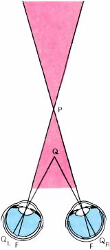
Rice. 113. Transection of the corpus callosum leads to loss of stereopsis in the shaded part of the visible space.
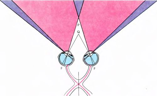
Rice. 114. Results of longitudinal transection of the chiasm along the midline. The subject will not see at all the two darker zones at the edges of the picture, on the left and on the right. Between these zones, where the color is lighter, there will be no stereopsis, with the exception of a small triangular-shaped zone behind point P (nothing is visible here at all) and a zone in front of point P (here stereopsis will remain).
If a person fixes his gaze on point P, then the projection of point Q, located closer to the eyes within the acute angle FPF, is QL and QR - will appear in the left and right eyes on opposite sides of the central fovea. Accordingly, the projection QL transmits information to the left hemisphere, and the Q projectionR - to the right hemisphere. In order to see that point Q is closer than P (i.e., to obtain a stereo effect), you need to combine information from the left and right hemispheres. But the only way to do this is to transmit information along the corpus callosum. If the path through the corpus callosum is destroyed, the person will be stereoblind in the area shaded in the figure. In 1970, D. Mitchell and K. Blakemore of the University of California, Berkeley, studied stereoscopic vision in one person with a transected corpus callosum and obtained exactly the result predicted above.
The second question, closely related to the first, is what kind of stereopsis disturbance will occur if the optic chiasm is cut along the midline (as R. Myers did on cats). The result here will be in a certain sense the opposite. From Fig. 114 it should be clear that in this case each eye will become blind with respect to stimuli falling on the nasal region of the retina, i.e. emanating from the temporal part of the visual field. Therefore, there will be no stereopsis in the lighter-colored area of space, where it is normally present. The lateral zones outside this area are generally accessible only to one eye, so there is no stereopsis here even under normal conditions, and after cutting the chiasm they will be zones of blindness (this is shown in a darker color in the figure). In the area behind the fixation point, where the temporal parts of the visual fields overlap, now invisible, blindness will also occur. However, in the area closer to the fixation point, the remaining hemifields of both eyes overlap, so stereopsis should be preserved here, unless the corpus callosum is damaged. K. Blakemore nevertheless found a patient with a complete transection of the chiasm in the midline (this patient, as a child, received a skull fracture while riding a bicycle, which apparently led to a longitudinal rupture of the chiasm). During the examination, he was found to have exactly the combination of vision defects that we have just hypothetically described.
Eye, brain, vision