Deprivation and development
Until now, we have imagined the brain as a fully formed, mature mechanism. We were interested in how its parts are connected to each other, how these parts function in everyday situations and how they serve the interests of the animal. All this, however, left open a completely different and extremely important question: how is this mechanism created?
There are two main components to this problem. Much of the brain development occurs before the animal is born, in the mother's womb. At first glance, the brain of a newborn baby, although it has fewer folds and is somewhat smaller than the brain of an adult, is otherwise not very different from it. A first superficial glance, however, is unlikely to reveal all the details; after all, a child, of course, is not born knowing the alphabet or with the ability to play tennis (or the harp). All these achievements involve learning, and by learning, of course, we mean the formation or modification of neural connections under the influence of the external environment. Thus, the final state of the brain is the result of both prenatal and postnatal development. First, this development includes maturation as such, determined by the internal properties of the organism and occurring before and after the moment of birth; secondly, it presupposes postnatal maturation, determined by training, education, education and experience - all these words are more or less equivalent.
Prenatal development is a gigantic topic; I don't know too much about it, so I won't describe it in any detail. One of the most interesting, but also the most difficult questions is how individual nerve fibers of a huge bundle find their destination. The eye, geniculate body and cortex, for example, form independently of each other; as they mature, the axons growing from them must make choices among many alternatives. The optic nerve fiber must grow through the retina to the blind spot, then pass as part of the optic nerve to the chiasm and make a decision here about whether to go to the opposite side; it must then proceed to the outer geniculate body of the chosen side, approach the desired layer (or an area that will later become the desired layer), and then exactly to the desired part of that layer so that the resulting topography becomes properly ordered; and finally, it must branch, and the branches must approach the appropriate parts of the geniculate body cell - its body or dendrite. Similar requirements apply to fibers growing from the lateral geniculate body to zone 17 or from zone 17 to zone 18. Although this general aspect of neuronal development is currently being intensively studied in many laboratories, we do not yet know how fibers find their destinations. It is difficult to even predict the winner among several major competing factors, which include mechanical induction, chemical gradients, and interactions with complementary molecules such as those occurring in the immune system. Numerous modern studies seem to point to the role of many different mechanisms.
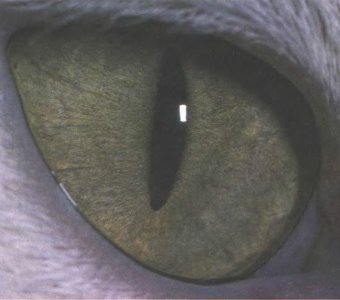
Rice. 132. Apparently, the slit shape of the pupil, as in this cat and in many nocturnal animals, makes it possible to reduce the intensity of light more effectively than a round pupil.
In this chapter we will look mainly at the postnatal development of the mammalian visual system, especially the influences that the system experiences from its environment. With respect to the first levels of the cat and monkey visual system—the retina, the geniculate body, and perhaps the striate (primary visual) cortex—a natural question arises as to whether some plasticity persists after birth. I'll start by describing a simple experiment. Around 1962, a number of important facts about the visual cortex of the adult cat became known: orientation selectivity was discovered, “simple” and “complex” cells were discovered, and many cortical neurons became known to be activated by signals from both eyes or to exhibit varying degrees of ocular dominance. We knew enough about the adult animal to ask direct questions about how plastic the visual system is. So Torsten Wiesel and I took a week-old kitten, when his eyes were about to open, and sewed his eyelids on one eye. This procedure may seem cruel, but it was carried out under anesthesia, and the awakened kitten, returned to its mother and other kittens, showed no signs of discomfort or distress. Ten weeks later, we surgically opened the eye, again under anesthesia, and began recording the responses of the cortical neurons to determine whether turning off the eye had any effect on the eye itself or on the visual system.
Before I describe the results, I should note that the idea for this experiment was inspired by a long history of psychological research and observations related to clinical neurology. Psychologists experimented extensively with visual deprivation on animals in the 40s and 50s, using behavioral methods to evaluate the results. A typical experiment involved keeping the animal in complete darkness from birth. When animals were brought into the light, they turned out to be blind or at least have defective vision. Blindness was somewhat reversible, but visual function was restored only gradually and usually not completely.
In parallel with these experiments, clinical observations were carried out on children with congenital cataracts. Cataract is a condition in which the lens of the eye becomes cloudy, allowing light to pass through but not allowing images to form on the retina. Cataracts in newborns, as in adults, are treated by surgical removal of the lens and implantation of an artificial lens or prescription of glasses with highly convex lenses. This allows you to restore a clear image on the retina. Despite the relative simplicity of the operation, ophthalmologists have been reluctant to perform it on very young children or newborns, mainly because any operation performed in early childhood is statistically associated with a large risk, although this risk is small. After cataract removal at, say, age eight, and glasses fitted, the results did not live up to expectations. Vision was not restored at all: the child, as before, remained blind, and deep defects persisted, despite months or years of attempts to “teach him to see.” The child, for example, still could not distinguish a circle from a triangle. As a result of the appearance and disappointment of hopes, the child’s condition usually worsened instead of improving. This is in sharp contrast to clinical cases with adults: a seventy-five-year-old man develops cataracts in both eyes and gradually loses his vision, but after three years of blindness the cataract is removed, glasses are fitted, and his vision is completely restored. Vision may even improve compared to what it was before the cataract developed, as the lens yellows with age and its removal results in the person again seeing the bright blue of the sky that only children and young people perceive.
Apparently, visual deprivation (deprivation of visual experience) in children leads to detrimental effects never seen in adults. Typically, psychologists quite reasonably explained the results of their experiments, as well as clinical results, by the child’s inability to “learn to see” or (which apparently amounts to the same thing) by the inability to form connections due to a lack of training experience.
Amblyopia - This is a partial or complete loss of vision, not associated with any anomalies of the eye itself. When we sutured the eye of a cat or monkey, our goal was to induce amblyopia and then figure out where in the optic tract the abnormality would occur. The results of the experiment with the kitten amazed us. Often an experiment produces inconclusive results that are only good enough to not completely discredit the idea, but not definitive enough to yield anything useful. However, our experiment was an exception: the results were completely clear - when we opened the kitten's eyelids, the eye itself looked completely normal; even the pupil contracted in the usual way when illuminated. However, the results of recording the activity of cortical cells could not be considered normal. Although we found many cells with completely normal responses to the orientation of the lines and their movement, we also found that, instead of about half the cells preferring one eye to the other, not one of the twenty-five cells examined was influenced in any way by the previously closed eye. (Five of the cells were uninfluenced by both eyes, something that is occasionally seen even in normal cats.) Compare this to a normal cat, in which about 15% of the cells are monocular, with about 7% responding to the left eye and 7% to the right. Histograms of eye dominance for a cat, shown at the top of Fig. 133, allow you to immediately see this difference. Undoubtedly, something has gone seriously wrong here.
We soon repeated this experiment on other kittens and baby monkeys. In kittens, a more extensive series of experiments soon showed that when the eyelids were sutured immediately after birth, on average, only 15 percent of the cells preferred the previously sutured eye, versus about 50 percent in intact kittens. Approximately the same results were obtained in monkeys (see the lower histograms in Fig. 133). Among the few cells that responded to stimulation of the previously closed eye, many appeared abnormal; they reacted sluggishly, quickly tired, and lacked precise orientation tuning.
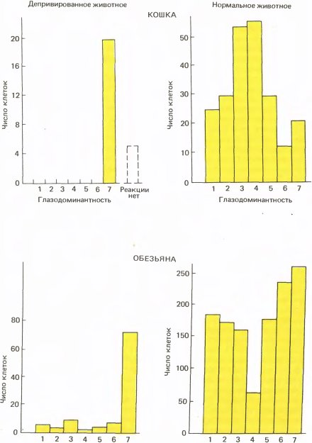
Rice. 133. The kitten's right eye was deprived of visual stimuli for two and a half months at about ten days of age, when the eyes are normally open. In this experiment (top histograms), we recorded responses from only twenty-five cells. (In subsequent experiments, we were able to examine more cells and found that a small proportion of them were influenced by the previously closed eye.) In experiments on baby monkeys (lower histograms), the results were very similar. His right eye was closed at two weeks of age and kept closed for 18 months. We later found that the same results were obtained by closing the eye for just a few weeks.
A result like this raises many questions. Where in the visual system did the anomaly occur? In the eye? In the cortex? Could the cat see with a previously closed eye, despite the cortical abnormality? Was this anomaly caused simply by light deprivation or by deprivation of the ability to see different shapes? Did it matter the age at which we closed our eyes? Was the anomaly the result of disuse of the eye or was it due to some other reason? It took a long time to figure all this out, but we can summarize the results in a few words.
In order to determine the location of the defect, it was obviously necessary to record reactions from lower levels, starting, say, from the retina or the geniculate body. The results were unambiguous: both in the retina and in the geniculate body there were many cells with almost normal reactions. The cells of those layers of the geniculate body to which fibers went from the previously closed eye had ordinary receptive fields with a center and periphery; they responded well to small spots and poorly to diffuse light. The only hint of an anomaly was a certain sluggishness in the reactions of these cells compared with the cells of the layers that received input signals from the normal eye.
Knowing this relative normality, we were amazed to see the Nissl-stained lateral geniculate body under the microscope for the first time. Its anomaly was visible practically even without a microscope. The geniculate body of a cat is organized somewhat more simply than that of a monkey; it consists mainly of two magnocellular layers located at the top, and not at the bottom, as in the monkey. The upper layer has input from the contralateral eye, and the lower layer has input from the ipsilateral eye. Beneath these layers lies a rather poorly defined layer of small cells with several subdivisions, which I am ignoring here. On each side, the magnocellular layer, which received inputs from the closed eye, was paler and clearly thinner than its partner, which looked completely normal. The cells of the abnormal layers were not only pale, but also shriveled to approximately two-thirds of their normal cross-section. The result obtained when closing the right eye is shown in Fig. 134. Similar results were obtained in macaques (Fig. 135). Thus, we were faced with a paradox that took several years to resolve: in the cells of the lateral geniculate body, which physiologically seemed relatively normal, histological examination showed obvious pathology. Be that as it may, we now had an answer to our original question, since the cortical cells, which showed virtually no response to stimulation of the previously closed eye, nevertheless apparently received a significant and seemingly normal flow of input signals from the geniculate body; this suggested that the primary damage was localized not in the eye or geniculate body, but mainly in the cortex. During histological examination of the cortex, we found absolutely no signs of anomaly. As we will see later, there were still anatomical defects in the cortex, but they were not revealed using the staining methods we used.
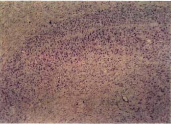
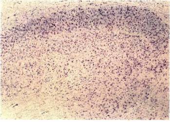
Rice. 134. If the right eye of a kitten is closed at the age of 10 days for three and a half months, then obvious anomalies will appear in its external geniculate bodies. Two main layers can be seen in the upper parts of both micrographs. Up: the upper layer on the left side (contralateral to the closed right eye) is thin and pale colored. Down: on the right (ipsilateral) side, the lower of the two layers is abnormal. Both layers together have a thickness of about 1 mm. The histogram of ocular dominance is shown in Fig. 133.
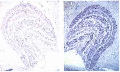
Rice. 135. Transverse sections of the left and right lateral geniculate bodies of a monkey whose right eye was closed at two weeks of age for 18 months. Abnormal layers are visible and appear paler. On both sides, these are the layers that receive input fibers from the previously closed (right) eye - layers 1, 4 and 6 on the left, 2, 3 and 5 on the right (counting from bottom to top).
The cells of the altered layers are smaller, but this cannot be seen at such a low magnification. The width of the entire structure is about 5 millimeters.
The next question was what exactly causes the anomaly when closing the eye. Stapling the eyelids reduces the amount of light reaching the retina by approximately 10 to 50 times; it, of course, also prevents the creation of any images on the retina. Could the anomaly be caused simply by a decrease in the amount of light? To find out, we inserted a frosted plastic contact lens with the consistency of a table tennis ball into one eye of a newborn kitten. In other animals, we instead sewed onto one eye a thin translucent membrane, acting as an extra eyelid, called the nictitating membrane, which is present in cats but absent in humans. The plastic or membrane reduced the amount of light by only half, but prevented the formation of any focused images. The results were the same: abnormal cortical physiology and abnormal geniculate histology. Apparently the damage was caused by an inability to perceive shapes, and not simply by a lack of light.
In several kittens, we tested vision before recording neural responses by placing an opaque black contact lens on the eye that was not covered and then observing the animal's behavior. The animals were undoubtedly blind to the deprived eye: being placed on a low table, they confidently walked to its edge, stepped over it and fell onto the bedding laid out on the floor. While walking on the floor, they usually bumped into table legs. No normal, self-respecting cat ever does this. Similar tests with the eye uncovered showed that vision was completely normal.
Next, we conducted long-term studies in cats and monkeys to determine whether the results depended on the age at which the eye was closed and the duration of deprivation. It soon became clear that age is critical. An adult cat in which one eye was disabled for more than a year showed no blindness in that eye, no loss of cortical responses, or pathology of the geniculate body. (The first deprived cat, the mother of our first litter of kittens, was an adult by definition!) After much experimentation, we concluded that there must be a period of plasticity somewhere between birth and adulthood when deprivation leads to a cortical defect. As it turns out, the cat has this critical period lasts from the fourth week to the fourth month. It is not surprising that closing the eye has little effect until the fourth week, since in the first month of life the cat uses almost no vision: the eyes open only around the 10th day, and for the next few weeks the kittens and their mother hide behind the sofa. Sensitivity to deprivation increases rapidly and reaches a maximum in the first weeks of the critical period. At this time, turning off one eye, even for several days, leads to a noticeable distortion of the ocular dominance histogram. Over the next four months, the time of deprivation required to produce obvious effects increases steadily; in other words, sensitivity to deprivation decreases and disappears.
In Fig. 136 shows histograms illustrating some of the results obtained in monkeys. The left graph shows the effect of turning off one eye for 6 weeks at the age of five days; there are almost no cells that respond to stimulation of a previously closed eye. A much shorter deprivation (middle graph) also causes a strongly pronounced effect, but clearly less than with longer deprivation. At the age of four months, sensitivity decreases so much that even turning off the eye for 5 years (right graph) gives an effect, although very noticeable, but still incomparable with the consequences of earlier deprivation.
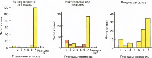
Rice. 136. Left: in a monkey whose right eye was sutured at the age of five days for six weeks, the left eye is almost completely dominant. In the middle: Closing the eye for just a few days at a few weeks of age is enough to cause a noticeable shift in ocular dominance. The darker color shows the number of abnormal cells. Right: If the closure of the eye is delayed until four months of age, then a very long shutdown (in this case for five years) leads to a much smaller shift in ocular dominance than a short shutdown at the age of several weeks.
When studying the period of sensitivity in cats and monkeys, very similar results were obtained. In monkeys, this period begins earlier, at birth rather than at four weeks of age, and lasts longer, gradually ending by one year rather than the fourth month. Sensitivity is highest in the first two weeks of life, during which just a few days of deprivation are sufficient to cause a pronounced shift in ocular dominance. Blackout of an eye in an adult monkey, regardless of its duration, does not cause any harmful effects. In one adult monkey we closed the eye for five years, after which there was no blindness, no cortical defect, no shrinkage of cells in the geniculate body.
Recovery
The next question that interested us was the possibility of any restoration of impaired function in a monkey after opening a previously disconnected eye. It turned out that no or almost no physiological recovery occurs if an eye, closed for a week or longer, is simply opened and nothing else is done. Even several years later, the cortex remained approximately the same abnormal as at the moment the eye opened (Fig. 137, left graph). If, having opened one eye, the other, previously open, was closed (we called this procedure reversion), then recovery was observed, but only if the monkey was still in the critical period (Fig. 137, middle and right graphs - for the case of early and late reversion). After the critical period ended, even reversion—closing the other eye for several years—provided little more than minor restoration of structure or function.
The monkey's ability to see did not always correspond to the physiological state of the visual cortex. Without reversal, vision in the previously closed eye was never restored. With reversal, vision returned and often reached an almost normal level, and this happened even in cases of late reversal when the physiology of the originally closed eye remained very abnormal. We are still unable to explain this discrepancy between the lack of significant physiological or anatomical normalization and the fact that in some cases vision appears to have been largely restored. It is possible that both observations reflect different aspects of visual function. We tested visual acuity by measuring things like the smallest discernible break in a line or circle. Such testing, however, may not provide a complete picture of visual function. It is difficult to believe that such pronounced physiological and anatomical defects manifest themselves at the behavioral level in just a slight decrease in visual acuity.
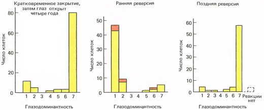
Rice. 137. Left: The macaque had one eye closed at birth for 9 days and then opened. Cell responses were recorded after four years, during which the animal's vision was tested repeatedly. Even such a long period when both eyes were open did not lead to a noticeable restoration of physiological parameters.
In the middle: the right eye has been disabled since birth; After five and a half weeks, this eye was opened and the left eye was closed. When responses were recorded in the right hemisphere at six months of age, the initially closed right eye strongly dominated the majority of cells.
Right: in this case, the right eye was closed at 7 days of age for one year, after which it was opened and the left eye was closed. After another year, the left eye was also opened, after which both eyes remained open. When cortical responses were finally recorded at the age of three and a half years, most cells responded to signals from the eye that was open at the very beginning. Obviously, one year is too late for a reversion.
Nature of damage
The results described above clearly show that the absence of retinal images in early life leads to profound and persistent impairments in cortical function. However, two important questions remained open regarding the nature of this phenomenon. The first was a question related to the nature versus nurture dilemma: were we depriving animals of the individual experiences they needed to form the right connections, or were we destroying the connections that were already there, pre-formed, and functioning when the animal was born? Almost all experiments with raising animals in the dark, conducted decades before our work, were interpreted in the context of the problem of learning. The cerebral cortex, which most people believed (and still believe) to be the physiological substrate of memory and thinking, was seen as something like the 1-megabyte storage device that we pay so much for when we buy a computer: both contain many elements and connections, but there is no information until we put it there. In short, the bark was considered a kind of tabula rasa.
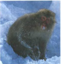
Rice. 138. Japanese macaque (Macaca fuscata), The largest of all macaques, it lives on the ground and in trees in northern Japan. It is protected from the cold by thick grayish-brown fur.
One obvious approach to solving this question would be to directly record neuronal activity in newborn kittens or monkeys. If learning were required to create the necessary connections, we would not find anything like the highly developed specificity observed in adult animals. The lack of specificity, however, would not solve the problem, since the underdevelopment of connections could be explained either by immaturity (the fact that the construction of a genetically programmed system of connections has not yet been completed) or by a lack of sensory experience. On the other hand, the discovery of such specificity would argue against a learning mechanism. We didn't expect the experiments on kittens to be simple; That's how it turned out. The kitten is born with a very immature visual system and does not use its eyes at all until around the tenth day when they open. At this time, even the media of the eye, the transparent substances located between the cornea and the retina, are not yet completely transparent and do not allow obtaining a clear image on the retina. The immature visual cortex did respond sluggishly, somewhat unpredictably, and was generally very different from the normal visual cortex of the adult cat; however, we found many cells with clear orientation specificity. The more time passed from birth to registration, the greater the number of cells in their behavior approached the cells of the adult type - perhaps due to the clarification of the ocular media and the general strengthening of the body, and perhaps as a result of learning. Different groups of researchers explain this in different ways.
The most convincing data were obtained in newborn monkeys. In macaques, the day after birth, the visual system displays amazing maturity: the babies look in different directions, follow objects with their gaze and show great interest in their surroundings (Fig. 139). In accordance with this behavior, the cells of the primary visual cortex in them exhibit the same acute orientation tuning as in the adult. Consistently located cells are well ordered in orientation (see graph in Fig. 140). Although we noted some differences between newborn and adult animals, the orientation-specific receptive field system, a distinctive feature of the striate cortex, seemed quite clearly organized.
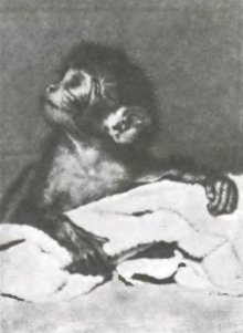
Rice. 139. The day after birth, the baby macaque looks around, fixes objects with his gaze, and shows a keen interest in the world around him. In humans and cats, visual function reaches this level of maturity only many weeks after birth.
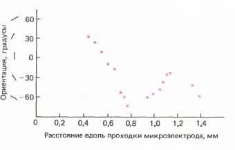
Rice. 140. In the newborn macaque, cortical cells exhibit approximately the same orientation selectivity as in adults, and the sequence of orientations is almost as ordered.
In the macaque (compared to the cat or human), the visual system of the newborn may be considered quite mature, but it is certainly anatomically different from the visual system of the adult monkey. Nissl-stained sections of the cortex look different: the young have thinner layers and a denser packing of cells. Le-Wey was the first to show that even the total area of the striate cortex increases by about 30 percent from birth to adulthood. If the cortex is stained using the Golgi method or examined under an electron microscope, the differences become even more obvious: the cells typically have less developed dendrites and fewer synapses. In view of these differences, it would be surprising if the cortex at birth functioned exactly the same as in an adult animal. On the other hand, a month after birth, dendrites and synaptic connections are still not fully developed. The question “nature or nurture?” means: is postnatal development determined by experience or still proceeds according to the innate program? We are not yet sure of the answer, but judging by the comparative normality of the reactions of cortical cells at birth, we can conclude that their disruption after deprivation is mainly due to the “damage” of connections that existed at the time of birth, and not the impossibility of their formation without individual experience.
The second important question concerns the cause of this damage. At first glance, the answer seemed almost obvious. We believed that the degradation of the connections is due to their lack of use, just as atrophy of the leg muscles occurs when the knee or ankle is fixed with a plaster cast. In the shrinkage of the cells of the geniculate body we saw a manifestation postsynaptic atrophy - this is the name for the decrease in the size of cells in the external geniculate bodies of adult animals or humans after removal of the eye. These assumptions turned out to be incorrect. We considered them so self-evident that I'm not sure we would have ever thought of doing a special experiment to test them. We had to change our point of view only as a result of one unnecessary, as it seemed to us then, experiment, the reasons for which I no longer remember.
We sutured both eyes first on a newborn kitten and then on a newborn baby monkey. If the failure of the responsiveness of cortical cells receiving input from one eye were due to their inactivity, then turning off both eyes would double the defect: we would be virtually unable to detect cells responding to the left or right eye. In fact, to our great amazement, we did not get anything like the absence of cellular responses: after opening the eyes, a good half of the cells in the striate cortex again responded normally, one quarter responded abnormally, and one quarter did not respond at all. We were forced to conclude that it was impossible to predict the fate of a cortical cell in a situation where one eye was closed if it was unknown whether the other was also closed. Disable one eye and the cell will almost certainly lose its connections to it; but turn off both eyes and you have a good chance of maintaining the connection. We are obviously faced here not with the inaction of cells and their connections, but with some kind of competition between the two eyes. The situation is as if the cell initially had two groups of synaptic inputs - two input paths, one from each eye, but if one path is not used, the other takes over, taking over the territory of the first (Fig. 141).
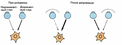
Rice. 141. We propose that the cortical cell has inputs from two sources, one from each eye, and that closing one eye weakens connections from that eye and strengthens connections from the other.
Such reasoning, we believed, is unlikely to apply to the shrinkage of cells of the geniculate body, since these cells are monocular and there is no visible possibility of competition here. At that time, we were unable to explain the atrophy of cells in the layers corresponding to the closed eye. When both eyes are closed, the shrinkage of the geniculate cells is not so noticeable, but it is difficult to say anything here, since there are no normal layers that could be used for comparison. Our understanding of this whole problem did not advance until we began to use some new methods of experimental anatomy.
Strabismus
The most common cause of amblyopia in people is strabismus, or strabismus - non-parallelism of the optical axes of the eyes. Strabismus can be convergent or divergent. The cause of strabismus is unknown, but there are almost certainly multiple causes. Sometimes strabismus occurs after birth, in the first months of life, when the eyes barely begin to fix and trace objects. Disturbances in directness of gaze may result from abnormalities in the eye muscles or brainstem mechanisms that support eye movement.
In some children, strabismus appears to be associated with farsightedness. To properly focus a distant object, the lens of a farsighted eye must take on the same convex shape as the lens of a normal eye when focusing a near object. Rounding of the lens when viewing close objects means contraction of the ciliary muscle inside the eye; this process is called accommodation. When a person with normal vision performs near-distance accommodation, the eyes simultaneously automatically rotate toward the median plane (convergence). These two processes are shown in Fig. 142. Brainstem neural networks governing accommodation and convergence are likely interconnected and possibly overlapping; be that as it may, it is difficult to carry out one of these reactions without the other. When a farsighted person makes the accommodation necessary to focus even a distant object, one or both eyes may turn inward, although convergence is not desirable in this case. If a farsighted child does not wear glasses, turning the eye may become a permanent habit over time. This explanation of strabismus is, of course, only true in some, but not all cases, since strabismus is not always accompanied by farsightedness, and in some people with strabismus one eye turns outward rather than inward.
Strabismus can be treated surgically by changing the attachment sites of the extrinsic eye muscles. Surgery is usually successful in correcting the alignment of the eye axis, but until the last decade it was most often postponed until the child reached the age of four to ten years (for the same reason that cataract removal was delayed - to somewhat reduce the risk of surgery).
Strabismus that occurs in adults, for example due to damage to the nerve or eye muscle, is, of course, accompanied by double vision. To experience this condition, you just need to apply light pressure on one eye from below and from the side. Double vision can be quite distressing and even incapacitating, and if no better solution is possible, one eye should be covered with a patch. Otherwise, double vision will remain until the squint is corrected. In children with strabismus, however, double vision rarely continues for a long time; it is replaced either by alternating use of both eyes, or by suppression of vision in one eye.
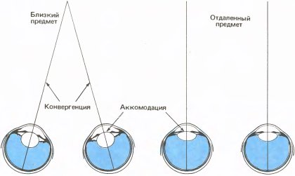
Rice. 142. When viewing a close object, two adaptive reactions occur: the lens is rounded as a result of contraction of the ciliary muscles, and the eyes turn to the median plane of the head.
When a child alternates eyes, he fixates objects first with one eye, while the non-fixating eye turns inward or outward, and then with the other eye, while the first eye moves to the side. (Alternating strabismus is very common, and if you know its signs, you can easily recognize it.) The eyes are used in turns, sometimes changing almost every second, and while one eye is looking, the other seems to see nothing. Any time one eye is positioned correctly and the other is deviated, vision is said to be in the deviated eye depressed. Suppression is familiar to anyone who is accustomed to looking through a monocular microscope, aiming a gun, or performing any other task using only one eye while the other eye is open. For a depressed eye, visual perception simply disappears. A child with alternating strabismus always suppresses one eye or the other, but if the vision in each eye is tested separately, both eyes will usually be found to be quite normal.
Some children with strabismus do not alternate eyes, but use one eye all the time while suppressing the other. If one eye is habitually suppressed, the vision in the suppressed eye tends to deteriorate. Visual acuity decreases, especially in and around the fovea, and if this condition lasts long enough, the eye can become virtually blind. Ophthalmologists call this type of blindness amblyopia ex anopsia. This is the most common type of amblyopia and even blindness in general.
Naturally, it occurred to us to try to induce strabismus and therefore amblyopia in kittens or monkeys by surgically cutting one of the eye muscles after birth, since by doing this we could carry out physiological studies and see how much of the visual pathway was impaired. We did this on half a dozen kittens and found to our displeasure that the kittens, like many children, developed alternating strabismus; they looked first with one eye and then with the other. Having tested each eye separately, we soon became convinced that vision was normal in both eyes. It became clear that we had failed to induce amblyopia, and we began to think about what to do next. We decided to record cellular reactions on one of the kittens, although we had absolutely no idea what could be found out in this way (research often involves “groping” searches). The results were completely unexpected. Studying cell by cell, we soon realized that something strange had happened to the brain: each cell responded completely normally, but only to stimulation of one eye. As the electrode moved across the cortex, there was a series of neurons activated by the left eye, then this series broke off and the other eye came into action. Contrary to what we observed after eye closure, neither eye was disadvantaged over the other in the overall balance of dominance. Sometimes binocular cells appeared in the transition zone, but, as can be seen from Fig. 143, the binocular cell fields in the entire population were about 20 percent instead of the normal 85 percent.
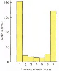
Rice. 143. If one of the outer eye muscles is cut in a newborn kitten, after three months the vast majority of the cells are monocular, falling into groups 1 and 7.
We became interested in what happened to the originally binocular cells - maybe they simply died or completely lost the ability to respond, so that only the primarily monocular cells remained? This seemed highly unlikely, since as the electrode advanced we found the usual abundance of responding cells - there was nothing like a fivefold decrease in their total number. In a normal cat, when the electrode is advanced horizontally in the upper layers of the cortex, we usually encounter about 10 to 15 cells in a row, dominated by the same eye; they all clearly belong to the same ocular dominance column, and two or three of them may be monocular. In cross-eyed kittens, we similarly observed 10–15 cells with dominance of one eye, but now all but two or three were monocular. Apparently, each cell completely or almost completely came under the jurisdiction of the eye that previously simply had a greater influence on it.
To appreciate the significance of this result, it is worth remembering that we essentially did not change the total number of visual stimuli received by each retina. Since we had no reason to believe that we had damaged either of the two eyes, we concluded that the overall flow of impulses in the two optic nerves should have remained normal.
How could strabismus cause such radical changes in cortical function? To answer this question, we need to look at how the two eyes interact with each other normally. Strabismus altered the relationship between stimuli affecting both eyes. When we look at an object, images of any point on it normally fall on those places of the two retinas that are at the same distance and in the same direction relative to both foveae - they fall on corresponding points. If an image on the left retina activates a binocular cell (this happens if the cell's receptive field is crossed by a dark/light boundary whose orientation exactly corresponds to that cell), then that cell will also be activated by an image on the right retina. This is due to three reasons:
1) images fall on the same areas of both retinas;
2) a binocular cell (unless it is specialized for depth perception) has receptive fields in exactly the same areas of both retinas;
3) the orientation preferences of binocular cells are always close for both eyes. Obviously, in the case of strabismus, reason 1 disappears: if, with mismatched images, one eye at a given moment induces the cell to discharge, then whether the other eye will do the same depends only on chance. For a single cell, this is apparently the only factor that changes during strabismus. When a kitten is in a situation for several weeks or months in which the signals from both eyes are no longer consistent with each other, this causes the weaker of the two sets of input connections of a given cell to become even weaker and often virtually disappear. Thus, we have here an example of pathological changes caused not by the absence of stimulation, but simply by a break in the normal temporal relationships between two sets of stimuli - a sophisticated blow, considering the seriousness of the consequences.
In similar experiments on monkeys, the same results were obtained; therefore, it appears that strabismus has the same effects in humans. According to clinical data, a person with long-term alternating strabismus, even after eliminating this defect, usually does not restore the ability to perceive depth. The surgeon can only align the two eyes to within a few degrees, and the patient has probably already lost the ability to achieve complete fusion of the two images by aligning the eyes correctly to within a few minutes of arc. Surgical correction of strabismus aligns the eyes well enough that normal neural mechanisms would handle the remaining few degrees of fine adjustment, but in strabismus it is these mechanisms, including the binocular cells of the cortex, that are disrupted. Recovery would likely require a long period of precise eye alignment, which involves both normal muscular adjustment and adjustment determined by binocular vision.
All these considerations explaining the shift in ocular dominance are very reminiscent of the model that allows one to explain associative learning by changes in synapses. This model was proposed by psychologist Donald Hebb of McGill University. Its basic idea is that the synapse between two neurons A and C becomes more effective the more often the excitation of neuron A is followed by the excitation of neuron C, regardless of what causes the latter (Fig. 144). Thus, to increase the efficiency of the synapse, it is not at all necessary that the discharge of neuron C be consequence discharge A. Suppose, for example, that another neuron, B, forms a synapse with C, and the synapse A–>C is weak, and the synapse B–>C is very effective; Let us further assume that A and B discharge almost simultaneously, or B slightly earlier than A, and that C discharges not under the influence of A, but as a result of the strong influence of B. In the Hebb model, the very fact that C discharges immediately after A increases the efficiency of the A->C synapse. We will also assume that if impulses coming to C from A are not accompanied by impulses to C, then the A–>C synapse weakens.
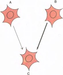
Rice. 144. Cell C receives input from left eye cell A and from right eye cell B. According to the Hebbian synapse model, if the discharges of cell C follow the discharges of cell A, then this sequence of events tends to strengthen the A->C synapse.
Let us apply this model to binocular convergence in a normal animal. Let cell C be binocular, axon A coming from the non-dominant eye, and axon B coming from the dominant eye. The non-dominant eye will activate cell C less often than the dominant eye. According to Hebb's hypothesis, the A->C synapse will persist or strengthen as long as impulses in A are followed by impulses in C, and such a sequence is more likely if, at the appropriate moment in time, “reinforcement” is constantly coming from the other eye along the B axon. And this is exactly what will happen when the images on both retinas are accurately combined. If activity in A is not accompanied by activity in C, the A–>C synapse will weaken over a long period of time. Obtaining direct evidence that the Hebbian model applies to strabismus will not be easy (at least in the near future), but I think the idea itself deserves attention.
Anatomical consequences of deprivation
That in the geniculate body, where there is little or no opportunity for eye competition, we could detect no noticeable physiological defects seemed to support the idea that the effects of closing one eye are due to competition rather than to ocular inactivity. True, the cells of the geniculate body were atrophic, but, we reasoned, one can hardly expect that everything will be clear. If competition really plays an important role, then cortical layer 4C might, it seemed to us, be a suitable object to test this idea, since the cells there are also monocular and competition is unlikely, so the alternation of stripes associated with the left and right eyes should remain undisturbed. So, by making long microelectrode penetrations through layer 4C, we began to find out whether such stripes persisted after the closure of one eye and whether their sizes remained normal. It soon became clear that layer 4C was still divided into sections of the left and right eyes, as in normal animals, and that the cells in the stripes connected to the previously closed eye remained essentially normal. However, the sequences of cells dominated by the closed eye were very short, as if the stripes were abnormally narrow (about 0.2 mm instead of 0.4 or 0.5 mm), and the stripes belonging to the open eye were correspondingly wider.
As soon as this became available, we began to use a technique that involved injecting a tracer into the eye and transneuronally transferring it to the cortex to obtain direct and visual confirmation of our conclusions. After a period of deprivation of several months, we injected the cat or monkey into one eye or the other with a radioactive amino acid. Autoradiographs showed a marked narrowing of the stripes associated with the previously deprived eye and a corresponding widening of the stripes belonging to the normal eye. Microphotograph on the left in Fig. 145 illustrates the results of injection of a radioactive amino acid into a normal eye. This image, taken under darkfield conditions as usual, shows a slice parallel to the surface of the cortex and passing through layer 4C. Narrow, interrupted black stripes correspond to the eye that was closed, and wider light (tagged) stripes correspond to the open eye into which the tag was injected. In Fig. 146 shows the opposite picture obtained after introducing a mark into a previously closed eye. This cut was made across layer 4C, so we see "streaks" in the cross section.
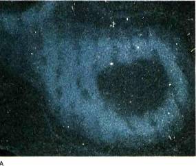
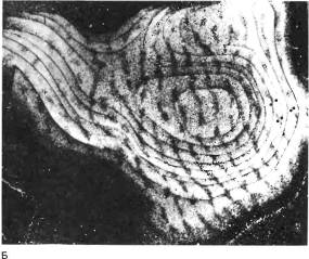
Rice. 145. Slices of the visual cortex of a macaque monkey whose right eye was closed from birth to 18 months of age. A radioactive amino acid was injected into the left eye, and brain slices parallel to the surface of the visual cortex were prepared a week later. The bark is dome-shaped, so such cuts first go tangentially and then form rings (A) increasingly larger in diameter, similar to onion rings. These rings were cut from microphotographs and glued together (B). Subsequently, we learned to straighten the bark before freezing it, and then we no longer had to cut and glue photographs of serial sections. On a typical autograph, the silver grains appear black on a white background. We, however, used a dark-field method in which silver grains scatter light and their clusters appear as bright areas. The light stripes—the places where labeled atoms enter the 4C layer from the open eye—are expanded, while the dark stripes (associated with the closed eye) are greatly narrowed.
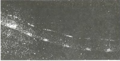
Rice. 146. Another monkey had a mark inserted into its closed eye. The cut is made in a transverse rather than tangential direction. In this dark-field image, the light streaks in layer 4C (seen here in cross-section) are greatly narrowed.
The results obtained on layer 4C confirmed our doubts about the competitive model associated with the fact of atrophy of the geniculate cells: either the competition hypothesis was incorrect, or there was some error in our reasoning. It turned out that the error concerned both the geniculate body and the cortex. In the cortex, our mistake was the assumption that by the time we closed the eyes of newborn animals, the ocular dominance columns were already fully formed.
Normal development of ocular dominance columns
An obvious way to obtain information about ocular dominance columns in newborn animals was to check the distribution of fibers included in layer 4C by injecting a tracer into the eye on the first or second day of life. The result was unexpected. Instead of clear, well-defined stripes in layer 4C, we observed a continuous distribution of label. Autoradiograph on the left in Fig. 147 shows this layer in cross-section, and we see no evidence of columns. Only if we cut the bark parallel to its surface could we see faint undulations at half-millimeter intervals, as shown in the autograph on the right. Apparently, the fibers growing from the geniculate body into the cortex do not branch immediately to the areas associated with the left and right eyes. They first send out their branches everywhere within a radius of a few millimeters and only later, around the time of birth of the animal, are they retracted and finally distributed. Slight waviness in a newborn indicates that retraction began before birth; Paško Rakic, with great difficulty, managed to inject the mark into the eye of a fetal monkey and found out that it begins several weeks before birth. By injecting a tracer into one eye at various points after birth, we were able to easily establish that in the first two or three weeks, a gradual retraction of the endings in layer 4 occurs, so that by the fourth week the formation of the stripes is complete. The pattern of the stripes and their periodicity in increments of 0.8 millimeters are thus innate.
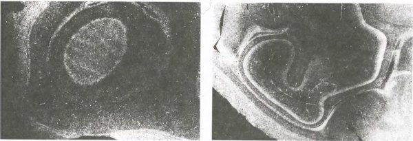
Rice. 147. Left: cross-section of layer 4C cortex of a newborn macaque after injection of a radioactive tracer into the eye. The photo was taken in a dark field, so the marked areas appear light. Their continuity shows that the inputs from each eye are not grouped into stripes, but are mixed throughout the layer. (The white stripe between layers 4C, near the surface of the brain and in the depths, represents the white matter of the brain, filled with labeled fibers coming from the lateral geniculate bodies.)
Right: here is a cross-section of the other hemisphere; When making a slice, the knife lightly touched the area of the striate cortex located in depth. You can see a hint of stripes at the top of layer 4C. (These stripes are located in the division associated with the magnocellular layers of the geniculate body.) A deeper sublayer? forms a continuous ring around ? and therefore presumably differentiates later by ocular dominance.
We easily confirmed the idea of postnatal terminal retraction by recording layer 4C responses from monkeys shortly after birth. As the electrode moved along this layer parallel to its surface, we were able to evoke activity from both eyes everywhere, instead of the clear alternation of eyes observed in adult animals. Carla Schatz demonstrated a similar development of the geniculate body in the cat: in the fetus, many cells of the geniculate body temporarily receive input from both eyes, but lose one of the inputs as a layered structure forms. Thus, in both the cortex and the geniculate body we have examples of the formation and subsequent spontaneous degeneration of synapses during development.
The final pattern of alternating stripes for the left and right eyes in cortex 4C develops normally even when both eyes are closed by suturing the eyelids; this means that proper connections can be created without the participation of individual experience. We believe that during development, inputs from both eyes compete in layer 4C in such a way that if inputs from one eye are dominant in some location, the advantage of that eye tends to increase, and the number of inputs from the other eye decreases accordingly. Even a small initial inequality tends to gradually increase until distinct stripes with complete dominance appear throughout layer 4 at the age of one month. When the eye is closed, the balance is disrupted, and at the boundaries of the stripes, where normally the outcome of the fight is decided by a direct collision, the open eye gains an advantage and gains the upper hand, as shown in Fig. 148.
We don't know what? during normal development it leads to an initial imbalance, but with such an unstable balance even the smallest difference can decide the matter. Why the developing pattern should consist of parallel strips, each 0.5 mm wide, is a matter of speculation. Some researchers suggest that axons from the same eye, when they come close, begin to attract each other, and from different eyes, they begin to repel each other, and at short distances, repulsion is weaker than attraction, so attraction wins. As the distance increases, the attractive force decreases faster than the repulsive force, so that at some distance repulsion wins. The width of the zone of these competitive interactions determines the size of the columns. According to mathematical calculations, to obtain parallel stripes (as opposed to a checkerboard pattern or islands of axons from the left eye in a solid mass of axons from the right eye) all that is required is that the boundaries between the columns be as short as possible. So we are able to explain the contraction and expansion of the columns by showing that while the eye was closed in early life, competition was still possible.
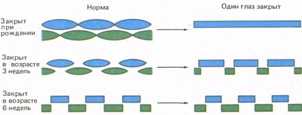
Rice. 148. This “competition model” explains the division of layer 4 fibers into columns of ocular dominance. By the time of birth, the columns had already begun to form. Normally, if at some point one eye dominates even slightly, this ends in its complete monopoly. If one eye is closed at birth, the remaining fibers from the open eye at any given point in layer 4 completely take over. Fibers from a closed eye are preserved only where they had no competitors at the time of closure.
Meanwhile, Ray Gillery, then at the University of Wisconsin, proposed a plausible explanation for cell atrophy in the geniculate body. Studying our drawings showing the shrinkage of cells after the closure of one eye in cats, he noticed that in the areas of the geniculate body most distant from the midline, the shrinkage was significantly less; Indeed, there - in the projection of the temporal crescent region - the cells seemed quite normal. Here the lateral edge of the visual field is displayed, so far from the middle that only one eye on this side can see it (Fig. 149). We were disappointed, to say the least, because we were so busy measuring cells to confirm our findings that we simply forgot to take a good look at our own micrographs. After all, the absence of atrophy of cells with inputs from the “temporal falx” meant that atrophy in other places of the geniculate body could, in fact, be the result of competition from the other eye.
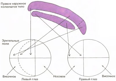
Rice. 149. Various areas of both retinas project to their own zones in the right geniculate body of the cat (which is shown here in cross section). The upper layer of the geniculate body, which receives input fibers from the contralateral (left) eye, overhangs the next layer. The pendulous part receives inputs from the temporal crescent of the visual field, which projects onto the retina of only one eye. When this eye is closed, the hanging part does not atrophy, presumably because it does not encounter competition from the other eye, which does not have the corresponding area.
Using a very ingenious experiment, shown in Fig. 150, Murray Sherman and colleagues provided decisive evidence for the role of competition in geniculate cell atrophy. First, they destroyed a tiny section of one retina in the kitten's binocular field of vision. They then stitched the other eye tightly shut. In a small area of the layer of the geniculate body onto which the eye with local damage was projected, severe atrophy was discovered. This result was also obtained by many other researchers. In the layer that had inputs from the other, previously closed eye, as expected, they also observed cell shrinkage everywhere except for the zone that had undergone atrophy in the layer with the projection of the other eye. There, despite the lack of input signals from the eye, the cells were normal. Atrophy due to eye closure was prevented by eliminating competition. It is clear that competition could not take place in the geniculate body itself; however, it should be remembered that although the cell bodies and dendrites of the geniculate body were located in the geniculate body itself, most of the endings of their axons were in the cortex, and there, as I have already said, the endings belonging to the closed eye were subject to severe atrophy. The conclusion is that when the eye closes, the shrinkage of cells is the result of a decrease in the number of axon terminals they support.
Rice. 150. In 1974, an experiment by Sherman, Gillery, Kaas, and Sanderson demonstrated the importance of competition for cell atrophy in the lateral geniculate body. If a small area of a kitten’s left retina is destroyed, an island of severe atrophy will form in the corresponding part of the upper layer of the right lateral geniculate body. If the right eye is then closed, as might be expected, the layer below undergoes atrophy, with the exception of the area lying directly below the atrophied area of the upper layer. This fact strongly supports the competitive nature of atrophy caused by eye closure.
The discovery that at birth fibers from both eyes were represented without any interruption throughout layer 4 was very useful, since it explained how competition at the synaptic level could be realized in a structure in which there seemed to be no opportunity for interaction between the eyes. Still, the problem may not be so simple. If the reason for changes in layer 4 is that there are conditions for competition in the first weeks after birth, then closing the eye at an age when the system is still plastic and the columns are already separated would not lead to changes. We closed the eye at five and a half weeks of age and only a year later introduced a tag into the other eye. The result was a clear narrowing and widening of the corresponding bands. This seems to indicate that, in addition to the differentiated retraction of endings, their germination into a new territory is also possible.
Other special deprivation experiments
In all the studies described so far, we closed one or both eyes or cut the extrinsic muscles of one of the eyes. Soon, many other experiments were carried out in many laboratories, including almost every conceivable type of visual deprivation. One of the first and most interesting experiments asked whether keeping an animal in conditions that allowed it to see stripes of only one orientation would lead to the loss of cells sensitive to all other orientations. In 1970, Colin Blakemore and J. F. Cooper of the University of Cambridge exposed kittens to alternating black and white vertical stripes for several hours every day from an early age, and kept them in the dark the rest of the time. The result was a preservation of cortical cells that responded to vertical stripes and a sharp decrease in the number of cells that preferred other orientations. It is unclear whether cells with initially intermediate orientations stopped responding altogether or whether they changed their preferred direction to vertical. In a paper published the same year, Helmut Hirsch and Nico Spinelli used glasses that allowed a kitten to see only vertical contours with one eye and only horizontal contours with the other. The result was a cortex containing cells with a preference for verticals, cells with a preference for horizontals, but very few cells with a preference for slanted lines. In addition, cells activated by horizontal lines were affected only by the eye that had previously been exposed to horizontal lines, and cells activated by vertical lines were affected only by the eye previously exposed to vertical lines.
Other interesting procedures included raising animals in a dark room in which a bright pulse of light was flashed one or more times per second; it allowed the animal to see where it was, but had to minimize the perception of any movement. The result of these experiments, which were carried out in 1975 by Max Zinader, Nancy Berman and Alan Hayne at the Massachusetts Institute of Technology, and by M. Zinader and G. Chernenko at Dalhousie (Halifax), was a decrease in the number of cells sensitive to movement. In another series of experiments, begun by F. Tretter, M. Zinader and Wolf Singer in Munich, animals were shown only the movement of stripes from left to right and the expected asymmetric distribution in the cortex of cells sensitive to the direction of movement was obtained. At great difficulty and expense, Torsten Wiesel and I raised baby monkeys in a room illuminated only by long-wavelength red light, and then recorded the responses of cells in the lateral geniculate body to see if there were unusually few color-coding neurons there (see Chapter 8). It was not possible to detect any anomalies in the geniculate bodies.
Finally, in the last decade, a lot of research has appeared to determine whether modifiable synapses contain special neurotransmitters or neuromodulators such as norepinephrine, acetylcholine or serotonin. The results of these studies will be of great interest.
The implications of deprivation outcomes in a broader perspective
It is often asked what purpose the plasticity of the visual system serves in early life (in humans, the period of sensitivity is supposed to be four to five years). In animals that have one eye closed from birth, the territory of the open eye in layer 4C expands; does this give any advantage to the open eye? There is no answer to this question yet. It is difficult to imagine that visual acuity becomes better than normal, since normal acuity, measured by an ophthalmologist using a test table, is ultimately determined by the density of receptor packing (see Chapter 3), and it is already limited by the wavelength of light.
Be that as it may, it seems very unlikely that plasticity was developed during evolution simply in case the baby might lose an eye or develop strabismus. A popular and certainly plausible idea is that plasticity provides the fine-tuning of connections necessary for the perception of shape, motion, and depth, and that this tuning occurs largely postnatally under the control of vision itself. This idea is attractive because the brain's ability to learn can relieve it of the need to program all the details in advance and can provide enough flexibility to adapt to a variety of environmental conditions. However, there is no convincing experimental evidence for such an idea yet. I am personally inclined to think that in the primary visual cortex, and perhaps at several subsequent levels, connections are entirely determined by genetic instructions. It is clear that a significant proportion of these connections are formed prenatally and, therefore, without the participation of sensory experience, and whatever strategy is used to build them could also fine-tune them.
I am not suggesting that the same is true in other areas of the cortex. Most neuroscientists believe that the neural circuits responsible for language acquisition are located primarily in the cortex—and no one would argue that we are born knowing the intricacies of our native language. The modifiability of various cortical areas and the age range within which modifications are possible can vary widely from area to area, with the primary visual cortex, with its minimal plasticity limited to the fewest years of postnatal development, apparently representing an extreme case. At more peripheral levels of the visual system, including the retina, geniculate nucleus, and primary cortex, plasticity may simply be a byproduct of maturation; Here, I believe, competition plays a certain role, and interference in its implementation leads to a distortion of connections. But we don't really yet know why the visual cortex might be modified early in life.
It is worth noting that all modifications in our experiments were the result of anomalous early experiences and led to the formation of anomalous connections. Experiments purporting to show that enriched early experiences lead to enhanced development of the cortex or other brain structures do not seem very convincing to me.
What amazed us most about the visual deprivation experiments was the ability to induce noticeable physiological and morphological changes in the nervous system without actual physical intervention. It has long been known that cells in the nervous system can degenerate if the nerve is physically cut or crushed, but in the experiments I described the entire effect was limited to the exclusion of light, and in the experiments with strabismus the intervention was even more subtle. In each case, the "punishment" more or less corresponds to the "crime". Eliminate form, and cells that normally respond only to forms will stop responding to them. Change the relative position of the eyes by cutting one of the muscles, and the connections that normally serve binocular interactions are severed. Eliminate moving stimuli altogether or moving them in a certain direction, and the corresponding cells will not respond.
It hardly takes much imagination to think that a child deprived of social contact, or an animal raised in isolation, as in some of Harry Harlow's experiments, might experience similar, equally real changes in the areas of the brain that determine emotions or interactions with other individuals of the same species. Of course, no pathologist has yet observed any changes, but even after visual deprivation, it is also impossible to see any changes in the cortex without very special methods (like injection of a mark and eyes). When some axons shorten and others lengthen, the entire structure, even when examined under an electron microscope, looks exactly the same.
Thus, the possible significance of deprivation experiments extends far beyond the visual system - it also concerns neuroscience in general and, to a large extent, psychiatry. Freud may well have been right in linking psychoneuroses with events in early childhood, and given his neurological training, I think he would have been delighted by the idea that such events could lead to noticeable histological or histochemical changes in the actual physical brain.
Eye, brain, vision